

Case Reports
doi: 10.4103/jpn.JPN_78_17.
Psammomatoid Juvenile Ossifying Fibroma: Report of Three Cases with a Review of Literature
Affiliations
- PMID: 29675079
- PMCID: PMC5890560
- DOI: 10.4103/jpn.JPN_78_17
Item in Clipboard
Case Reports
Psammomatoid Juvenile Ossifying Fibroma: Report of Three Cases with a Review of Literature
J Pediatr Neurosci.
2017 Oct-Dec.
Abstract
Psammomatoid juvenile ossifying fibroma (PJOF), a variant of juvenile ossifying fibroma (JOF), is a locally aggressive neoplasm of the children and young adults. This entity has predilection for the sinonasal region. It forms a differential diagnosis for many bone neoplasms. We report three cases of PJOF, in young patients whose biopsy showed the presence of psammomatoid bodies in a cellular fibrous stroma. The diagnosis of JOF indicates requirement of extensive surgery due to its locally aggressive nature.
Keywords: Aggressive; juvenile; psammomatoid.
Conflict of interest statement
There are no conflicts of interest.
Figures
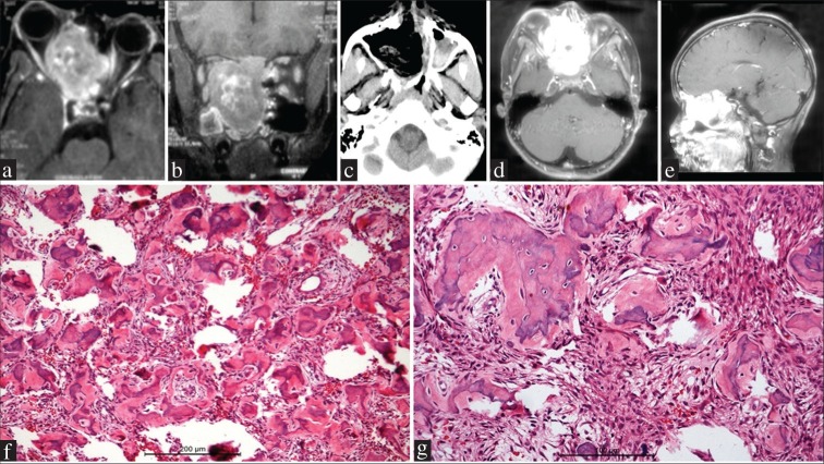
Case 1. (a and b) Magnetic resonance imaging brain postcontrast axial (a) and coronal (b) sections showing a large tumor occupying the sphenoid and ethmoid sinuses, extending into the medial wall of the right orbit and displacing the globe. The tumor enhances well on contrast and is extending into the anterior cranial fossa base and filling the nasal cavity (c) Postoperative computed tomographic scan showing near-total resection of the tumor. (d and e) Magnetic resonance imaging brain postcontrast axial (c) and sagittal (d) sections performed 1 year later showing regrowth of the contrast enhancing tumor involving paranasal sinuses and nasal cavity, with anterior cranial fossa base infiltration (f and g) Spherical calcified ossicles resembling psammoma bodies are embedded in a cellular fibrous stroma. The ossicles have irregular margins in contrast to psammoma bodies and are variably calcified (f = H and E, ×200; g = H and E, ×400)
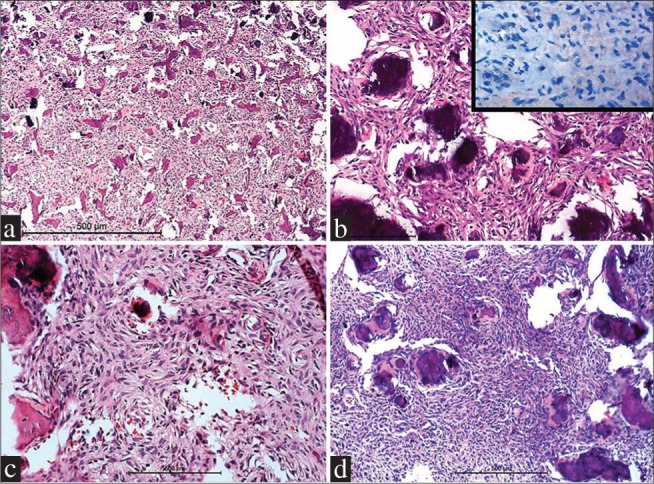
Case 2 (a and b) and Case 3 (c and d) showing basophilic psammomatoid ossicles in a cellular background of fibroblastic tissue. Tumor cells are negative for epithelial membrane antigen (inset, B) (a = H and E, ×100; b = H and E, ×400, c and d = H and E, ×200)
References
-
- Brannon RB, Fowler CB. Benign fibro-osseous lesions: A review of current concepts. Adv Anat Pathol. 2001;8:126–43. - PubMed
-
- El-Mofty S. Psammomatoid and trabecular juvenile ossifying fibroma of the craniofacial skeleton: Two distinct clinicopathologic entities. Oral Surg Oral Med Oral Pathol Oral Radiol Endod. 2002;93:296–304. - PubMed
-
- Solomon M, Khandelwal S, Raghu A, Carnelio S. Psammomatoid juvenile ossifying fibroma of the mandible: A histochemical insight. Internet J Dent Sci. 2009;7:2.
-
- Sarode SC, Sarode GS, Waknis P, Patil A, Jashika M. Juvenile psammomatoid ossifying fibroma: A review. Oral Oncol. 2011;47:1110–6. - PubMed
Publication types
LinkOut - more resources
Full Text Sources
Other Literature Sources
