Review
. 2017 Dec 18;9(4):790-810.
doi: 10.1039/c7sc04004k. eCollection 2018 Jan 28.

Advances in targeting the folate receptor in the treatment/imaging of cancers

Affiliations
Review

Advances in targeting the folate receptor in the treatment/imaging of cancers

Marcos Fernández et al. Chem Sci. .

Abstract

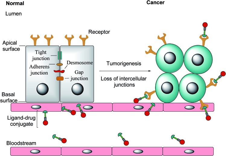
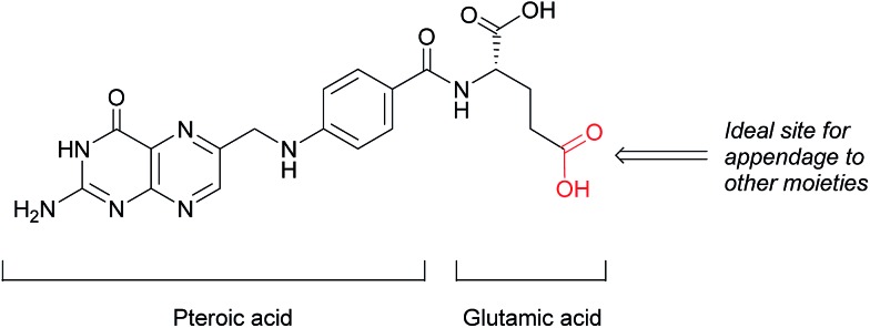
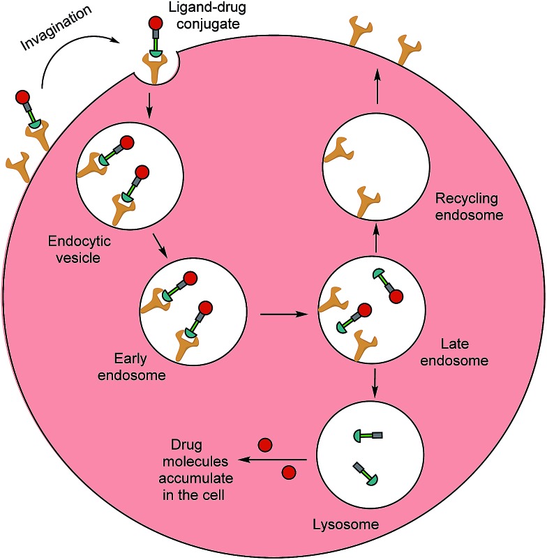
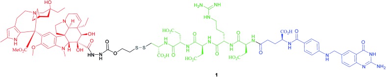
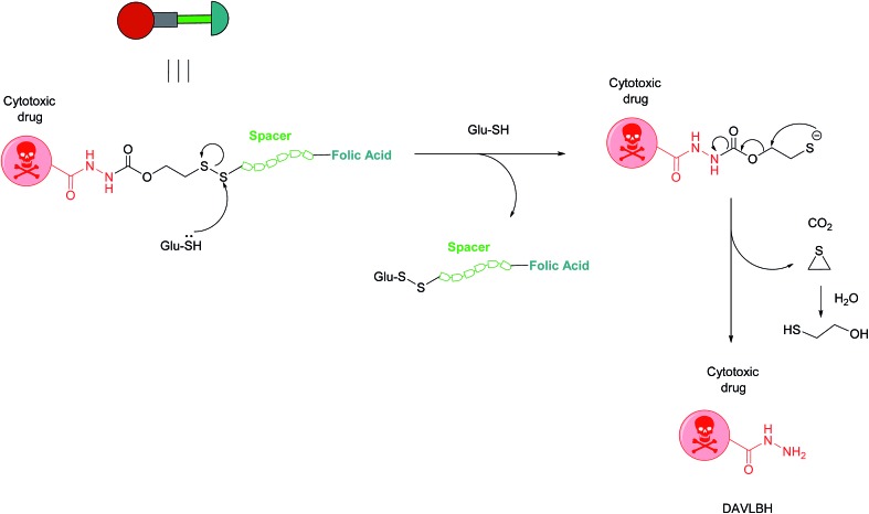
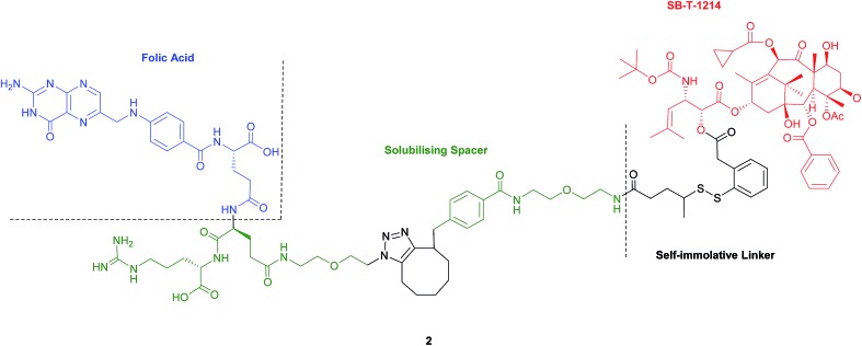
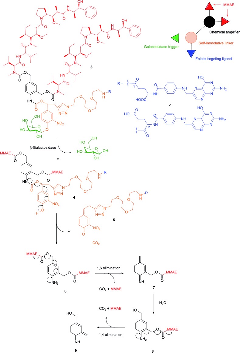
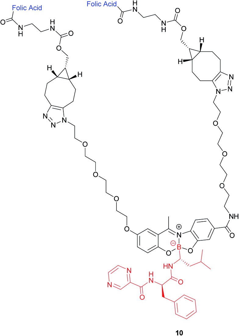
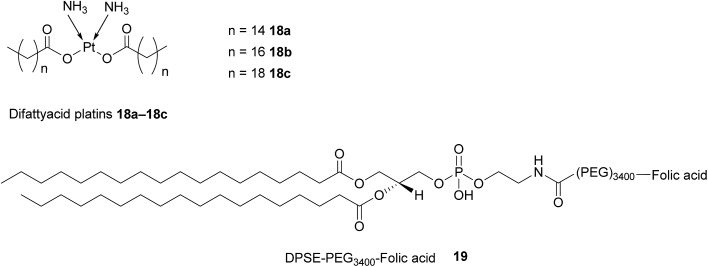
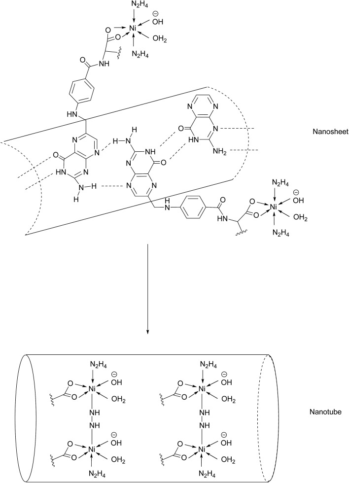
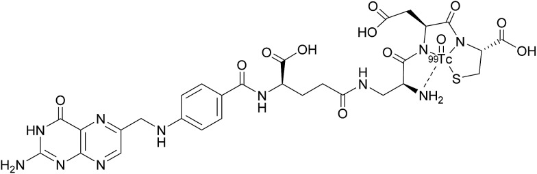
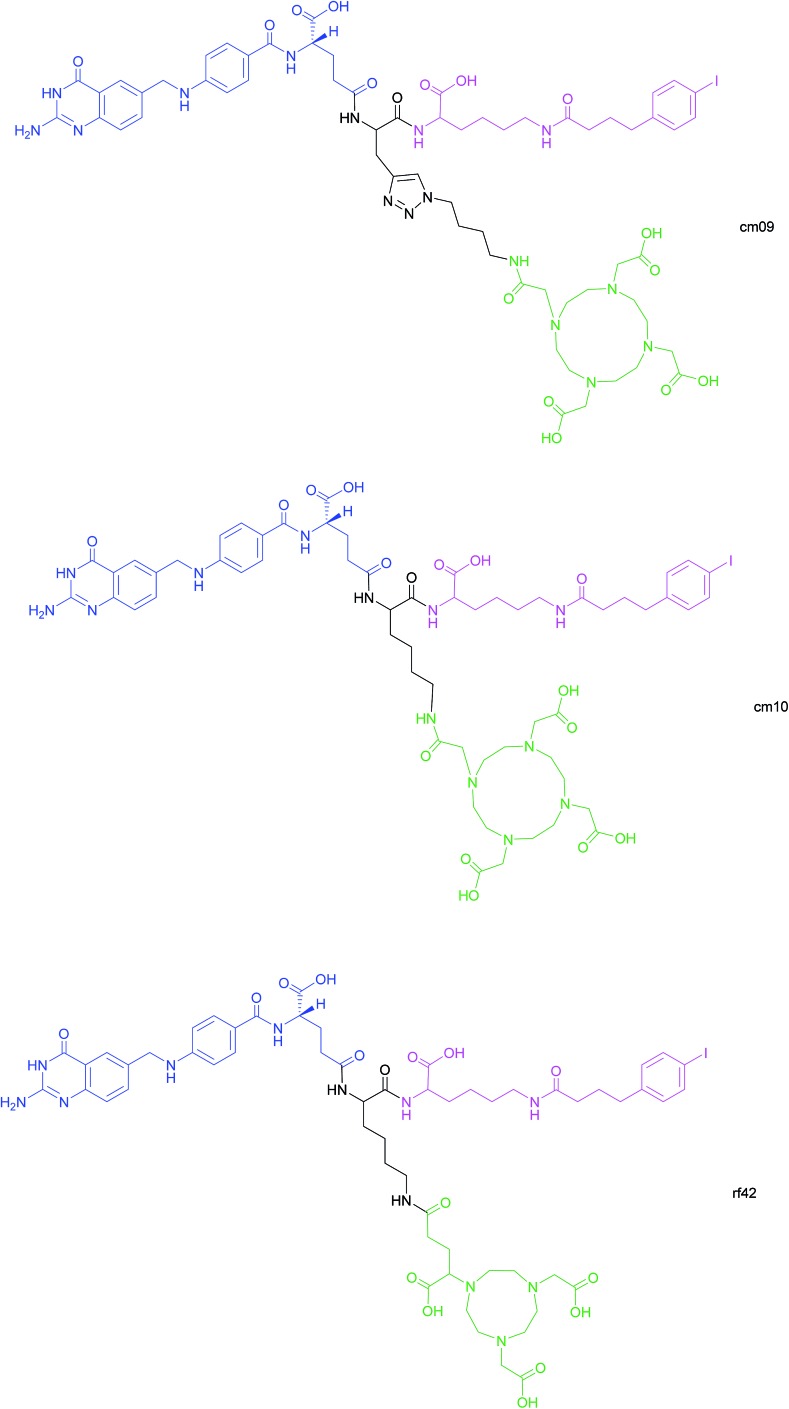
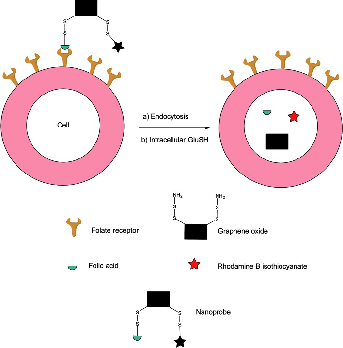
The folate receptor (FR) is a recognised biomarker for tumour cells due to its overexpression on a large number of tumours. Consequently, the FR has been exploited by many diagnostic and therapeutic tools to allow targeted delivery to, and imaging of, cancer cells. Herein, we describe the many different approaches by which this has been achieved, including the attachment of folate to potent chemotherapeutic drugs to form FR-targeting small molecule-drug conjugates (SMDCs), FR-targeting antibodies (as antibody alone and as an antibody-drug conjugate), and in the form of complementary nanotechnology-folate platforms; as well as imaging variants thereof. The potential of exploiting the FR for targeted therapy/imaging has the potential to revolutionise the way several cancers are treated. These FR-targeted technologies can also pave the way for inspiring further sophisticated drug conjugates, especially as this receptor is being targeted by use of several complementary technologies: small molecule, nanoparticle and protein-based - thus providing broad and distinct knowledge in the area.


Figures

Fig. 1
Fig. 1. The transformation of healthy epithelial cells to tumour cells and the effect it has on receptor positioning. Upon tumorigenesis, intercellular junctions (tight junctions, adherens junction, desmosomes and gap junctions) are lost and receptors that were previously only found on the apical surface become randomly positioned on the tumour.
Fig. 2
Fig. 2. Structure of folic acid (FA). This vitamin's constituent units are pteroic and glutamic acid.
Fig. 3
Fig. 3. Receptor-mediated endocytosis of a folate–drug conjugate. The construct initially binds to FRα or FRβ, forming an invagination and enclosing the conjugate in the early endosome. A mild drop in pH alters the receptor's conformation, resulting in FA–drug detachment. The late endosome's subsequent fusion with the lysosome leads to degradation of the conjugate and release of the free cytotoxic drug into the cell. The recycling endosome delivers the folate receptors back to the cell surface.
Fig. 4
Fig. 4. Chemical structure of the folic acid-based SMDC vintafolide 1 is comprised of a folate targeting ligand (blue), a peptide spacer (green), a self-immolative disulfide linker (grey) and the potent cytotoxic drug DAVLBH (red).
Fig. 5
Fig. 5. Mechanism of vintafolide drug release in the intracellular milieu. Glutathione cleaves the disulfide linker and the resulting thiolate undergoes a self-immolative, 1,2-elimination reaction to liberate the free drug DAVLBH.
Fig. 6
Fig. 6. Structure of the folate–taxoid conjugate 2 developed by Seitz et al.
Fig. 7
Fig. 7. Enzyme-catalysed double drug release mechanism of β-galactosidase-responsive folate–MMAE conjugate 3.
Fig. 8
Fig. 8. Structure of the boron complex 10 developed by Gois et al. consisting of (i) a folic acid targeting moiety (blue), (ii) PEG chains and (iii) the cytotoxic agent bortezomib (red).
Fig. 9
Fig. 9. Proposed mechanism for GluSH-mediated release of bortezomib (15) from complex 11.
Fig. 10
Fig. 10. Chemical structure of folate-BODIPY conjugates 16a and 16b.
Fig. 11
Fig. 11. Structure of IMGN853 (17). The anti-FRα antibody is conjugated to the DM4 drug via a self-immolative disulfide linker.
Fig. 12
Fig. 12. Schematic representation of a NMI-350 nanoemulsion. Difattyacid platins and C6-ceramide are encapsulated in the lipid core and lapidated gadolinium and folate are attached to the surface.
Fig. 13
Fig. 13. Structure of the difattyacid cisplatins 18a–18c and the DPSE-PEG3400-FA spacer 19.
Fig. 14
Fig. 14. Nanotube formation from nanosheets.
Fig. 15
Fig. 15. Chemical structure of 99mTc-etarfolatide.
Fig. 16
Fig. 16. Chemical structures of albumin-binding folate conjugates cm09, cm10 and rf42. They are each composed of three modules: (i) folic acid for FRα-expressing tumour targeting (blue), (ii) an albumin-binding moiety (pink), and (iii) the chelator (green) (DOTA for cm09 and cm10 and NODAGA for rf42) for coordination of 68Ga or 64Cu. The three functionalities are connected via a lysine linker in radiofolates cm10 and rf42, and by a triazole linker in the case of conjugate cm09.,,
Fig. 17
Fig. 17. Fluorescence off-on response of nanoprobe in FRα-positive cell.
None
Marcos Fernández
None
Faiza Javaid
None
Vijay Chudasama

Similar articles

Cited by

References

    1. Seitz J. D., Vineberg J. G., Herlihy E., Park B., Melief E., Ojima I. Bioorg. Med. Chem. 2015;23:2187–2194. - PMC - PubMed
    1. Krall N., Pretto F., Decurtins W., Bernardes G. J. L., Supuran C. T., Neri D. Angew. Chem., Int. Ed. 2014;53:4231–4235. - PubMed
    1. Valeur E., Knerr L., Ölwegård-Halvarsson M., Lemurell M. Drug Discovery Today. 2017;22:841–847. - PubMed
    1. Kularatne S. A., Wang K., Santhapuram H. K., Low P. S. Mol. Pharm. 2009;6:780–789. - PubMed
    1. Bhuniya S., Maiti S., Kim E. J., Lee H., Sessler J. L., Hong K. S., Kim J. S. Angew. Chem., Int. Ed. 2014;53:4469–4474. - PubMed

LinkOut - more resources