. 2017 Dec 6;9(5):1173-1183.
doi: 10.1039/c7sc04416j. eCollection 2018 Feb 7.

Glutaredoxins employ parallel monothiol-dithiol mechanisms to catalyze thiol-disulfide exchanges with protein disulfides

Affiliations

Glutaredoxins employ parallel monothiol-dithiol mechanisms to catalyze thiol-disulfide exchanges with protein disulfides

Ashwinie A Ukuwela et al. Chem Sci. .

Abstract

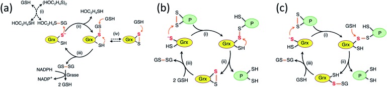
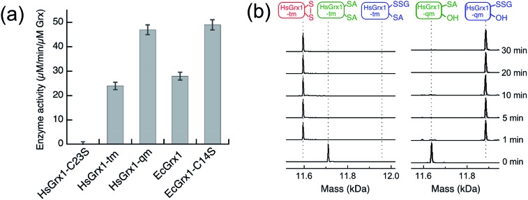
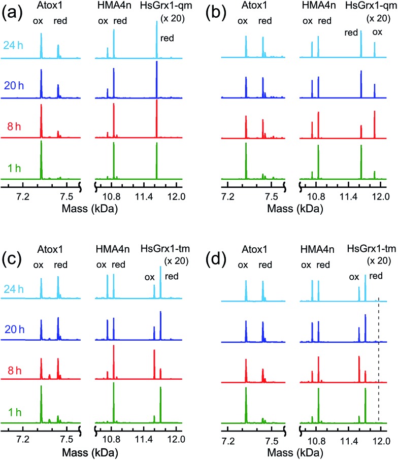
Glutaredoxins (Grxs) are a family of glutathione (GSH)-dependent thiol-disulfide oxidoreductases. They feature GSH-binding sites that directly connect the reversible redox chemistry of protein thiols to the abundant cellular nonprotein thiol pool GSSG/GSH. This work studied the pathways for oxidation of protein dithiols P(SH)2 and reduction of protein disulfides P(SS) catalyzed by Homo sapiens HsGrx1 and Escherichia coli EcGrx1. The metal-binding domain HMA4n(SH)2 was chosen as substrate as it contains a solvent-exposed CysCys motif. Quenching of the reactions with excess iodoacetamide followed by protein speciation analysis via ESI-MS allowed interception and characterization of both substrate and enzyme intermediates. The enzymes shuttle between three catalytically-competent forms (Grx(SH)(S-), Grx(SH)(SSG) and Grx(SS)) and employ conserved parallel monothiol and dithiol mechanisms. Experiments with dithiol and monothiol versions of both Grx enzymes demonstrate which monothiol (plus GSSG or GSH) or dithiol pathways dominate a specific oxidation or reduction reaction. Grxs are shown to be a class of versatile enzymes with diverse catalytic functions that are driven by specific interactions with GSSG/GSH.


Figures

Fig. 1
Fig. 1. Protein molecular structures. (a) Fully reduced HsGrx1 (PDB: 1JHB; thioredoxin fold); (b) HsGrx1(C8,26,79,83S)–GSH complex (; 1B4Q); (c) reduced EcGrx1 (; 1EGR); (d) Zn(ii)-AtHMA4n (; 2KKH; ferredoxin fold); (e) Cu(i)-Atox1 (; 1TL4). Labelled amino acid residues and the GSH fragment are shown as sticks while the metal ions in Zn(ii)-AtHMA4n and Cu(i)-Atox1 are represented as spheres.
Scheme 1
Scheme 1. (a) Proposed chemical pre-equilibrium monothiol mechanism for the classic bis(2-hydroxyethyl)disulfide (HEDS) assay., It consists of two coupled enzyme reactions employing HEDS as the disulfide substrate; (b and c) proposed dithiol mechanism (b) and monothiol mechanism (c) for catalytic reduction of protein disulfide P(SS) by Grx/GSH.
Fig. 2
Fig. 2. Protein speciation and reaction progress analysis upon oxidation of HMA4n(SH)2 (10 μM) in deoxygenated KPi buffer (50 mM, pH 7.0) containing GSSG (400 μM)/GSH (40 μM): IAA/ESI-MS analysis and speciation distribution for non-catalytic oxidation (a and b) and for catalysis by HsGrx1-tm (100 nM; c and d) and by HsGrx1-qm (50 nM; e and f). RS-A refers to the alkylated thiol RS-CH2CONH2.
Fig. 3
Fig. 3. (a) Comparison of substrate turnover rates for catalytic oxidation of HMA4n(SH)2 by different Grx enzymes under the condition of Fig. 2; (b) IAA/ESI-MS analysis for HsGrx1-tm (left panel) and HsGrx1-qm (right panel) during the catalysis of Fig. 2. RS-A refers to the alkylated thiol RS-CH2CONH2.
Fig. 4
Fig. 4. Reduction of HMA4n(SS) by GSH in KPi buffer (50 mM, pH 7.0, 100 mM NaCl): (a) IAA/ESI-MS analysis of HMA4n (10 μM) in the buffer containing GSSG (20 μM)/GSH (800 μM) without a Grx enzyme, inset: expanded view for the two minor HMA4n components; (b) the same analysis upon addition of HsGrx1-tm (100 nM) into (a); (c) reduction time course with no enzyme control and with either HsGrx1-tm or HsGrx1-qm as catalyst (each 100 nM); (d) correlation of catalytic rate with HsGrx1 enzyme concentrations (note: the catalytic rates given in Table 1 are obtained from the slopes of the best linear fits of the first three data points with enzyme concentration ≤ 0.1 μM).
Fig. 5
Fig. 5. IAA/ESI-MS analysis of reaction progress and protein speciation for thiol–disulfide exchange between Atox1(SS) and HMA4n(SH)2 (each 10 μM) in deoxygenated Mops buffer (50 mM, pH 7.0) with either monothiol or dithiol Grx enzymes: (a) monothiol HsGrx1-qm (0.5 μM) (indistinguishable from the result with no enzyme present); (b) monothiol HsGrx1-qm (0.5 μM) plus GSH (1.0 μM); (c) dithiol HsGrx1-tm (0.5 μM); (d) dithiol HsGrx1-tm (0.5 μM) plus GSH (1.0 μM). Note: for protein dithiol, the oxidized (ox) and the reduced (red) form is P(SS) and P(SA)2, respectively and for protein monothiol, the oxidized (ox) and the reduced (red) form is P(SSG) and P(SA), respectively; the dashed line in (d) indicates the position for the putative species HsGrx1-tm(SA)(SSG).
Scheme 2
Scheme 2. Dithiol mechanism in the absence of GSH (a) and monothiol mechanism in the presence of GSH (b) employed by Grx enzymes catalyzing thiol–disulfide exchange between proteins P1(SS) and P2(SH)2.
Scheme 3
Scheme 3. Proposed parallel monothiol–dithiol mechanism for catalytic oxidation of protein dithiol P(SH)2 (such as HMA4n(SH)2) by Grx/GSSG. The monothiol oxidation route is shown in solid arrows whereas the dithiol oxidation route in dashed arrows. Under the oxidative conditions, the resting enzyme forms for dithiol Grx and monothiol Grx are Grx(SS) and Grx(SSG), respectively.
Scheme 4
Scheme 4. Proposed parallel monothiol–dithiol mechanisms for catalytic reduction of protein disulfides P(SS) by Grx/GSH. The monothiol reduction route is shown in solid arrows with the dithiol route in dashed arrows. Under the reducing conditions, the resting forms for dithiol and monothiol Grx enzymes are Grx(SH)(S) and Grx(OH)(S), respectively.

Similar articles

Cited by

References

    1. Dalle-Donne I., Colombo G., Gagliano N., Colombo R., Giustarini D., Rossi R., Milzani A. Free Radical Res. 2011;45:3–15. - PubMed
    1. Allen E. M., Mieyal J. J. Antioxid. Redox Signaling. 2012;17:1748–1763. - PMC - PubMed
    1. Ströher E., Millar A. H. Biochem. J. 2012;446:333–348. - PubMed
    1. Go Y. M., Jones D. P. Crit. Rev. Biochem. Mol. Biol. 2013;48:173–181. - PMC - PubMed
    1. Hanschmann E. M., Godoy J. R., Berndt C., Hudemann C., Lillig C. H. Antioxid. Redox Signaling. 2013;19:1539–1605. - PMC - PubMed

LinkOut - more resources