Review
. 2017 Jul 3:3:17032.
doi: 10.1038/cddiscovery.2017.32. eCollection 2017.

Caspases in retinal ganglion cell death and axon regeneration

Affiliations
Review

Caspases in retinal ganglion cell death and axon regeneration

Chloe N Thomas et al. Cell Death Discov. .

Abstract

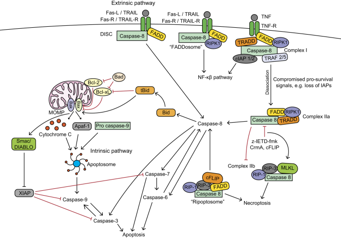
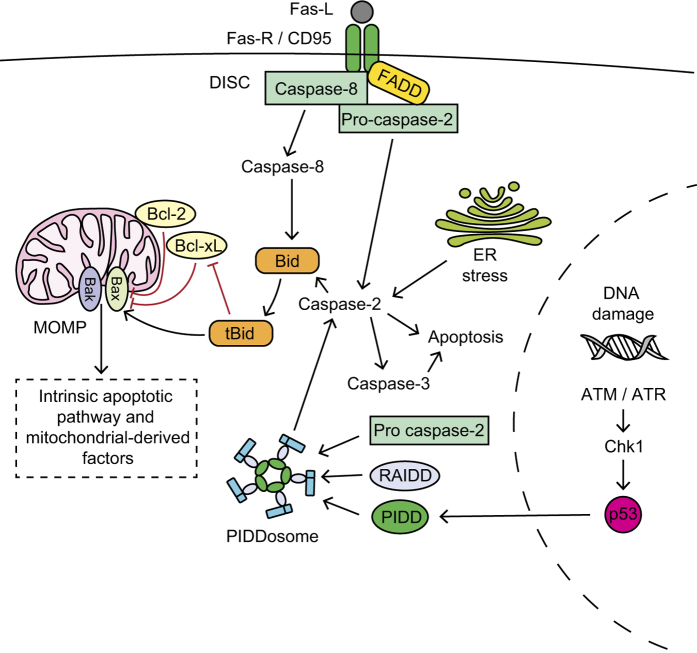
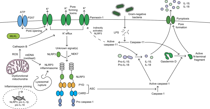
Retinal ganglion cells (RGC) are terminally differentiated CNS neurons that possess limited endogenous regenerative capacity after injury and thus RGC death causes permanent visual loss. RGC die by caspase-dependent mechanisms, including apoptosis, during development, after ocular injury and in progressive degenerative diseases of the eye and optic nerve, such as glaucoma, anterior ischemic optic neuropathy, diabetic retinopathy and multiple sclerosis. Inhibition of caspases through genetic or pharmacological approaches can arrest the apoptotic cascade and protect a proportion of RGC. Novel findings have also highlighted a pyroptotic role of inflammatory caspases in RGC death. In this review, we discuss the molecular signalling mechanisms of apoptotic and inflammatory caspase responses in RGC specifically, their involvement in RGC degeneration and explore their potential as therapeutic targets.


Conflict of interest statement

The authors declare no conflict of interest.

Figures

Figure 1
Figure 1
Apoptotic caspases in the canonical intrinsic and extrinsic pathways. Death receptor activation mediates the extrinsic pathway. Fas-R and TRAIL-R recruit FADD , and pro-caspase-8, forming the DISC, , leading to proximity-induced caspase-8 activation , and downstream activation of executioner caspase-3, -6 and -7. Caspase-8 can also activate the intrinsic pathway through truncating BH3-interacting domain death agonist (Bid) into tBid, which then promotes Bak and Bax mitochondrial membrane insertion, increasing MOMP and releasing apoptogenic factors, including Apaf-1, Cytochrome C and second mitochondria-derived activator of caspase/direct inhibitor of apoptosis-binding protein with low pI (Smac/DIABLO). , Cytochrome C, Apaf-1 and pro-caspase-9 form the septameric apoptosome complex, , which activates caspase-9 and successively downstream executioner caspases. Smac/DIABLO indirectly promotes apoptosis by opposing XIAP inhibition of caspase-3, -7 and -9. Caspase-8 can also form complex I at the TNF receptor, which upregulates the NF-κB survival inflammatory pathway; however, if survival signals are compromised (for example, IAPs) then complex I dissociates from the receptor forming complex IIa, which initiates caspase-8-dependent apoptosis. Caspase-8 inhibits complex IIb formation and necroptosis and caspase-8 inhibition (for example, through z-IETD-fmk) induces complex IIb formation, causing necroptosis. The ‘ripoptosome’ complex forms after cellular IAPs (cIAPs) or XIAP inhibition, causing caspase-8-dependent apoptosis and necroptosis. ,
Figure 2
Figure 2
Activation mechanisms of caspase-2. Caspase-2 is activated through DNA damage, upregulation of p53 and formation of the PIDDosome protein complex, which includes p53-induced protein with death domain (PIDD), RIP-associated ICH-1 homologous protein with death domain (RAIDD) and pro-caspase-2. , Caspase-2 is also activated by endoplasmic reticulum (ER) stress and at the Fas-R within the DISC, alongside Fas-associated protein with death domain (FADD) and caspase-8. Active caspase-2 cleaves and activates caspase-3, cleaves BH3 interacting domain death agonist (Bid; which initiates MOMP and the intrinsic apoptotic pathway) or initiates apoptosis directly.
Figure 3
Figure 3
Inflammatory caspase-1 is activated within the inflammasome protein complex; ,, which typically consists of a Nod-like receptor (NLR; such as Nod-like-receptor pyrin domain containing 3 (NLRP3)), adaptor protein apoptosis-associated speck-like protein containing a CARD (ASC) and caspase-1. Initial inflammasome priming is required for transcriptional upregulation of inflammasome machinery, such as NLRP3, pro-IL-1β and pro-IL-18. , A second signal then induces inflammasome assembly and activation. The NLRP3 inflammasome is activated by lysosomal rupture, reactive oxygen species (ROS), oxidised mitochondrial DNA (mtDNA) and cathepsin B. , Potassium (K+) efflux is a common NLRP3-activation mechanism, induced by P2X7-mediated pore opening, pore-forming toxins, pannexin-1 or MLKL-mediated pore opening. The NLRP3 inflammasome activates caspase-1, which cleaves precursor cytokines IL-1β and IL-18 into their active forms and gasdermin-D into its N-terminal fragment. The N-terminal fragment of gasdermin-D forms a plasma membrane pore facilitating pro-inflammatory cytokines release and inducing pyroptosis. Gram-negative bacterial lipopolysaccharide (LPS) can activate caspase-11, which also cleaves gasdermin-D cleavage and indirectly activates the NLRP3 inflammasome via pannexin-1.

References

    1. Berry M , Ahmed Z , Lorber B , Douglas M , Logan A . Regeneration of axons in the visual system. Restor Neurol Neurosci 2008; 26: 147–174. - PubMed
    1. Nicholson DW . Caspase structure, proteolytic substrates, and function during apoptotic cell death. Cell Death Differ 1999; 6: 1028–1042. - PubMed
    1. Nicholson DW , Thornberry NA . Caspases: killer proteases. Trends Biochem Sci 1997; 22: 299–306. - PubMed
    1. Jimenez Fernandez D , Lamkanfi M . Inflammatory caspases: key regulators of inflammation and cell death. Biol Chem 2015; 396: 193–203. - PubMed
    1. Fan T-J , Han L-H , Cong R-S , Liang J . Caspase family proteases and apoptosis. Acta Biochim Biophys Sin (Shanghai) 2005; 37: 719–727. - PubMed