

Differences of the Morphology of Subaxial Cervical Spine Endplates between Chinese and White Men and Women
- PMID: 29675423
- PMCID: PMC5838464
- DOI: 10.1155/2018/2854175
Differences of the Morphology of Subaxial Cervical Spine Endplates between Chinese and White Men and Women
Abstract
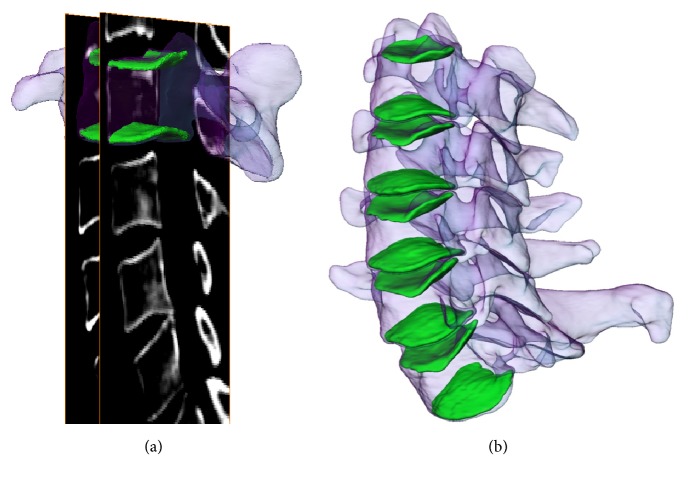
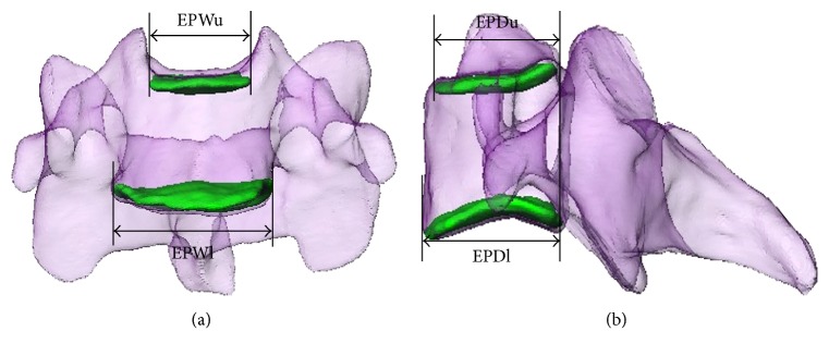
Objective. The aim of this comparative anatomical study was to specifically investigate endplate morphology differences between Chinese and White men and women. Materials and Methods. Three-dimensional cervical endplate models were constructed using computed tomography imaging of 41 healthy Chinese and 24 White subjects. The morphologic measurements of cervical endplate included linear parameters (EPWu: upper endplate width; EPDu: upper endplate depth; EPWl: lower endplate width; and EPDl: lower endplate depth) and area parameters with a digital measuring system. Results. All linear parameters showed a constant increase from C3 to C7 except for EPDl in both the Chinese and the White subjects. An increase trend was observed on area parameters in both Chinese and White subjects. The ratio of EPWl/EPDl was smaller in Chinese females than in White females at C3, C4, and C6 levels (P < 0.05). The ratio of EPWl/EPDl was significantly different between the Chinese and White men at C4-5 levels (P < 0.05). Conclusions. Our data indicates that the morphology of subaxial cervical spine endplates between Chinese and White men and women is different in most of the linear and area parameters. This information could provide guidelines for the design of CDA implants and the improvement of surgical techniques.
Figures

Similar articles
-
Quantitative anatomy of the endplate of the middle and lower cervical vertebrae in Koreans.Spine (Phila Pa 1976). 2007 Jun 15;32(14):E376-81. doi: 10.1097/BRS.0b013e318067e384. Spine (Phila Pa 1976). 2007. PMID: 17572609
-
Morphologic evaluation of Chinese cervical endplate and uncinate process by three-dimensional computed tomography reconstructions for helping design cervical disc prosthesis.J Chin Med Assoc. 2016 Sep;79(9):500-6. doi: 10.1016/j.jcma.2016.04.003. Epub 2016 May 25. J Chin Med Assoc. 2016. PMID: 27236369
-
A morphometric study of the middle and lower cervical vertebral endplates and their components.Medicine (Baltimore). 2017 Mar;96(10):e6296. doi: 10.1097/MD.0000000000006296. Medicine (Baltimore). 2017. PMID: 28272256 Free PMC article.
-
Kinematics of the subaxial cervical spine in rotation in vivo three-dimensional analysis.Spine (Phila Pa 1976). 2004 Dec 15;29(24):2826-31. doi: 10.1097/01.brs.0000147806.31675.6b. Spine (Phila Pa 1976). 2004. PMID: 15599286
-
Geometry of inferior endplates of the cervical spine.Clin Neurol Neurosurg. 2016 Mar;142:132-136. doi: 10.1016/j.clineuro.2016.01.027. Epub 2016 Jan 25. Clin Neurol Neurosurg. 2016. PMID: 26852320
Cited by
-
Effects of endplate coverage and intervertebral height change on heterotopic ossification following cervical disc replacement.J Orthop Surg Res. 2021 Nov 25;16(1):693. doi: 10.1186/s13018-021-02840-5. J Orthop Surg Res. 2021. PMID: 34823557 Free PMC article.
-
Does the sizing of current cervical disc arthroplasty systems match Chinese cervical anatomic dimensions?Front Bioeng Biotechnol. 2022 Oct 25;10:1036223. doi: 10.3389/fbioe.2022.1036223. eCollection 2022. Front Bioeng Biotechnol. 2022. PMID: 36394034 Free PMC article.
-
Reliability and reproducibility of measurements in para-sagittal planes on sub-axial cervical vertebral bodies: a morphometric study of endplates in three-dimensional models.J Orthop Surg Res. 2021 Aug 16;16(1):503. doi: 10.1186/s13018-021-02648-3. J Orthop Surg Res. 2021. PMID: 34399792 Free PMC article.
-
Sagittal morphometry of intervertebral spaces in subaxial cervical region of asymptomatic Chinese.Eur Spine J. 2024 Oct;33(10):3933-3940. doi: 10.1007/s00586-024-08462-9. Epub 2024 Aug 29. Eur Spine J. 2024. PMID: 39198288
-
The footprint mismatch of cervical disc arthroplasty comes from degenerative factor besides ethnic factor.Sci Rep. 2024 Sep 5;14(1):20673. doi: 10.1038/s41598-024-71786-5. Sci Rep. 2024. PMID: 39237767 Free PMC article.
References
-
- Kasliwal M. K., Traynelis V. C. Motion preservation in cervical spine: review. Journal of Neurosurgical Sciences. 2012;56(1):13–25. - PubMed
-
- Lin C.-Y., Kang H., Rouleau J. P., Hollister S. J., La Marca F. Stress analysis of the interface between cervical vertebrae end plates and the Bryan, Prestige LP, and ProDisc-C cervical disc prostheses: an in vivo image-based finite element study. The Spine Journal. 2009;34(15):1554–1560. doi: 10.1097/BRS.0b013e3181aa643b. - DOI - PubMed
-
- Richards O., Choi D., Timothy J. Cervical arthroplasty: the beginning, the middle, the end? British Journal of Neurosurgery. 2012;26(1):p. 2. - PubMed
Publication types
MeSH terms
LinkOut - more resources
Full Text Sources
Other Literature Sources
Medical
Miscellaneous
