

A case report of Ggeneralized uterine arteriovenous malformation after molar pregnancy in an infertile woman
- PMID: 29675497
- PMCID: PMC5899827
A case report of Ggeneralized uterine arteriovenous malformation after molar pregnancy in an infertile woman
Abstract
Background: Uterine arteriovenous malformation (UAVM) is a rare vascular condition in reproductive age presented mostly with bleeding. Although this malformation is infrequent, it is potentially life-threatening. Transvaginal Doppler ultrasonography is a widely available, noninvasive and excellent diagnostic method.
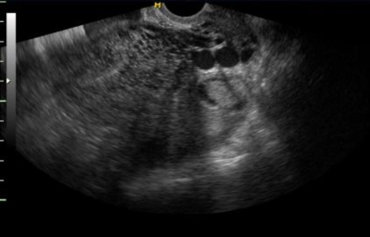
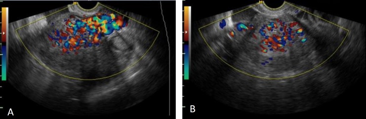
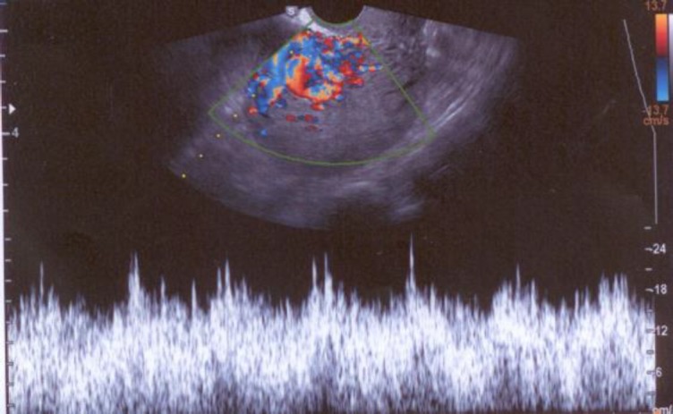
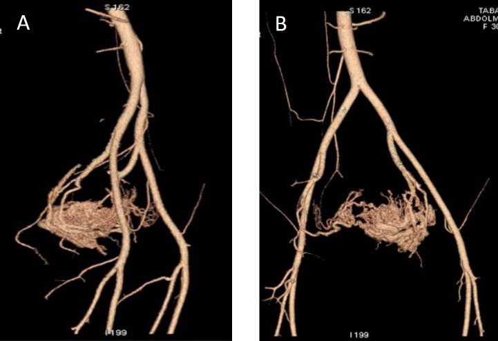
Case: The case is a 30-yr-old woman with a history of eight-yr infertility.following intrauterine insemination treatment, she had a molar pregnancy. Despite methotrexate treatment, there was persistent vaginal bleeding. Assessment of this patient was done with transvaginal sonography and color Doppler. According to suspicious appearances, angiography was planned for confirmation of UAVM.
Conclusion: UAVM is one of the molar pregnancy complications. The first step for diagnosis of UAVM is transvaginal ultrasonography and color Doppler assessment. Embolization is the best treatment for women who intend to preserve fertility.
Keywords: Arteriovenous malformation; Color doppler ultrasonography; Embolization; Molar pregnancy.
Conflict of interest statement
The author declares that he has no competing interests.
Figures
Similar articles
-
Ultrasound diagnosis and management of acquired uterine enhanced myometrial vascularity/arteriovenous malformations.Am J Obstet Gynecol. 2016 Jun;214(6):731.e1-731.e10. doi: 10.1016/j.ajog.2015.12.024. Epub 2016 Feb 9. Am J Obstet Gynecol. 2016. PMID: 26873276
-
Uterine arteriovenous malformation with repeated vaginal bleeding after dilatation and curettage.Obstet Gynecol Sci. 2019 Mar;62(2):142-145. doi: 10.5468/ogs.2019.62.2.142. Epub 2019 Mar 4. Obstet Gynecol Sci. 2019. PMID: 30918884 Free PMC article.
-
Uterine arteriovenous malformation (UAVM) as a rare cause of postpartum hemorrhage (PPH): a literature review.Arch Gynecol Obstet. 2022 Dec;306(6):1873-1884. doi: 10.1007/s00404-022-06498-0. Epub 2022 Mar 13. Arch Gynecol Obstet. 2022. PMID: 35284958 Review.
-
Fertility outcomes following pelvic embolization in women with acquired uterine arteriovenous malformation.Taiwan J Obstet Gynecol. 2017 Dec;56(6):831-835. doi: 10.1016/j.tjog.2017.10.023. Taiwan J Obstet Gynecol. 2017. PMID: 29241929
-
Pregnancy after uterine arteriovenous malformation-case series and literature review.Australas J Ultrasound Med. 2012 Aug;15(3):87-96. doi: 10.1002/j.2205-0140.2012.tb00012.x. Epub 2015 Dec 31. Australas J Ultrasound Med. 2012. PMID: 28191151 Free PMC article. Review.
Cited by
-
Uterine arteriovenous malformations as a rare differential diagnosis of abnormal uterine bleeding: A case report.Int J Surg Case Rep. 2025 Feb;127:110900. doi: 10.1016/j.ijscr.2025.110900. Epub 2025 Jan 16. Int J Surg Case Rep. 2025. PMID: 39826314 Free PMC article.
-
Uterine Arteriovenous Malformation: Approach and Treatment in a Nulligravidous Virgin Woman.Cureus. 2025 Jan 11;17(1):e77287. doi: 10.7759/cureus.77287. eCollection 2025 Jan. Cureus. 2025. PMID: 39931595 Free PMC article.
-
A Comparative Analysis of Follicular Diameter Assessment Versus Doppler Ultrasound in Predicting Ovulation Timing for the Infertility Treatment: Insights From a Prospective Study.Cureus. 2024 May 31;16(5):e61466. doi: 10.7759/cureus.61466. eCollection 2024 May. Cureus. 2024. PMID: 38953072 Free PMC article.
-
Congenital uterine arteriovenous malformation presenting as vaginal bleeding following vaginal delivery in a 23-year-old woman: A case report.Case Rep Womens Health. 2023 Mar 2;37:e00493. doi: 10.1016/j.crwh.2023.e00493. eCollection 2023 Mar. Case Rep Womens Health. 2023. PMID: 36915294 Free PMC article.
References
-
- Belfort P, Braga A, Freire NS. Malformação arteriovenosa uterine após doença trofoblástica gestacional. Rev Bras Ginecol Obstet. 2006;28:112–121.
-
- Fleming H, Ostor AG, Pickel H, Fortune DW. Arteriovenous malformations of the uterus. Obstet Gynaecol. 1989;73:209–214. - PubMed
-
- Shaaban AM, Menias CO, Tubay MS, Rezvani M, arouk El Sayed RF, Woodward PJ. Diagnostic imaging gynecology. 2nd Ed. Canada: Elseiver; 2015. pp. 2–163.
-
- O’Brien P, Nevastam A, Buckley AR, Chang SD, Legiehn GM. Uterine arteriovenous malformations from diagnosis to treatment. J Ultrasound Med. 2006;25:1387–1392. - PubMed
-
- Bottomoley JP, Whitehouse GH. Congenital arteriovenous malformation of the uterus demonstrated by angiography. Acta Radiological Diagnostica. 1975;16:43–48. - PubMed
Publication types
LinkOut - more resources
Full Text Sources