Review
. 2018 Aug;14(4):463-483.
doi: 10.1007/s12015-018-9802-4.

Pluripotent Stem Cells for Retinal Tissue Engineering: Current Status and Future Prospects

Affiliations
Review

Pluripotent Stem Cells for Retinal Tissue Engineering: Current Status and Future Prospects

Ratnesh Singh et al. Stem Cell Rev Rep. 2018 Aug.

Abstract

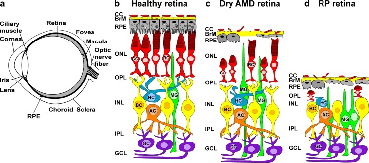
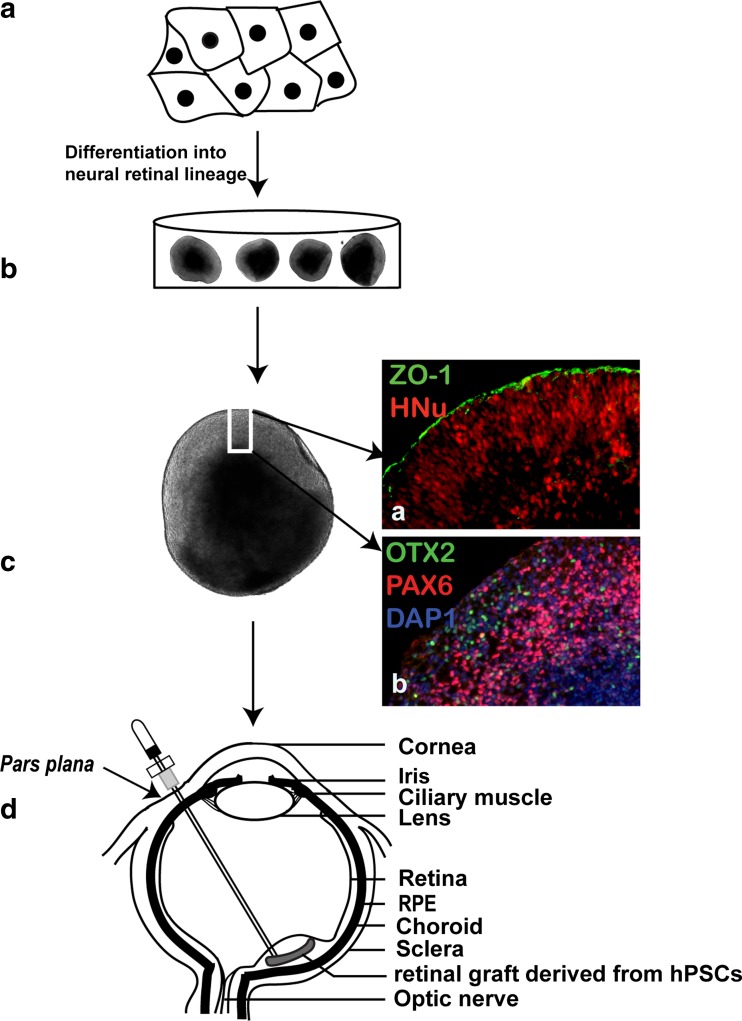
The retina is a very fine and layered neural tissue, which vitally depends on the preservation of cells, structure, connectivity and vasculature to maintain vision. There is an urgent need to find technical and biological solutions to major challenges associated with functional replacement of retinal cells. The major unmet challenges include generating sufficient numbers of specific cell types, achieving functional integration of transplanted cells, especially photoreceptors, and surgical delivery of retinal cells or tissue without triggering immune responses, inflammation and/or remodeling. The advances of regenerative medicine enabled generation of three-dimensional tissues (organoids), partially recreating the anatomical structure, biological complexity and physiology of several tissues, which are important targets for stem cell replacement therapies. Derivation of retinal tissue in a dish creates new opportunities for cell replacement therapies of blindness and addresses the need to preserve retinal architecture to restore vision. Retinal cell therapies aimed at preserving and improving vision have achieved many improvements in the past ten years. Retinal organoid technologies provide a number of solutions to technical and biological challenges associated with functional replacement of retinal cells to achieve long-term vision restoration. Our review summarizes the progress in cell therapies of retina, with focus on human pluripotent stem cell-derived retinal tissue, and critically evaluates the potential of retinal organoid approaches to solve a major unmet clinical need-retinal repair and vision restoration in conditions caused by retinal degeneration and traumatic ocular injuries. We also analyze obstacles in commercialization of retinal organoid technology for clinical application.


Conflict of interest statement

Authors do not have conflict of interest.

Figures

Fig. 1
Fig. 1
(a) Schematic drawing of a cross-section through a human eye. Light enters the eye through the cornea, passes through the pupil, lens and strikes the retina. Retina is the light-sensitive tissue lining the inner surface of the eye. Visual information from retina transmits to the brain through optic nerve fiber. In the middle of the retina there is a small depression called fovea, which is responsible for high-resolution vision. Region surrounding the fovea is called macula and is rich in cones. Retinal pigment epithelium (RPE) is a pigmented cell layer separating the choroidal blood supply from the photoreceptor layer. Choroid is a vascular layer of the eye. Sclera is a tough white sheath around the outside of the eye ball. (b) Schematic diagram of healthy retinal circuits. Mammalian retina consists of six major types of neuronal cells – rod cells (RC) and cone cells (CC), horizontal cells (HC), bipolar cells (BC), amacrine cells (AC) and retinal ganglion cells (RGC). The Müller cell (MC) are the glial cell that span across the retina and their somata. RPE provides metabolic and transport functions essential for homeostasis of the neural retina. Bruch’s membrane (BrM) is a highly specialized and multi-laminar structure separating RPE from the choroid and mediates exchange of oxygen and nutrients between vasculature of choroid and neural retina. RPE and the Bruch’s membrane form the outer blood–retinal barrier. Choroidal capillaries (CC) are the blood capillaries present in choroid that supply oxygen and nourishment to the outer layer of the retina. (c) Schematic diagram of dry age-related macular degeneration (AMD) retinal circuit. In Dry AMD, there is progressive atrophy of retinal pigment epithelium (RPE), Bruch’s membrane (BrM) and choriocapillaris (CC) in the macula. As a result, RPE cells stop providing support functions and the photoreceptors in the macula die, resulting in a loss of central vision. (d) Schematic diagram of retinitis pigmentosa (RP) retinal circuit: In RP, rod photoreceptors die, which trigger dramatic changes in the morphology of second order neurons (horizontal cells, bipolar cells and amacrine cells). As a result of the rapid rod degeneration, rod-driven bipolar and horizontal cell axon terminals retract their fine dendrites, and rod bipolar cell axon terminals assume immature synaptic structures. Defects extend to the cone circuit during the late phase of degeneration. In this case, both cones and cone horizontal cells sprout new neurites, whereas cone bipolar cells retract their dendrites
Fig. 2
Fig. 2
Schematic diagram showing transvitreal grafting of retinal tissue derived from hPSCs in subretinal space. (a) human pluripotent stem cell differentiated into retinal lineage. (b) Three dimensional retinal organoids growing in a dish. (c) Phase contrast image of retinal organoids showing different layers differentiating within retinal organoid. (a) hESCs derived retinal organoid coimmunostained for RPE marker ZO-1 and human nuclei marker HNu. (b) Retinal organoid coimmunostained for multipotential retinal progenitor markers OTX2 and PAX6. DAPI stained nuclei. d. A piece of retinal tissue derived from hESCs is transplanted transvitreally into subretinal space

References

    1. Martin GR. Isolation of a pluripotent cell line from early mouse embryos cultured in medium conditioned by teratocarcinoma stem cells. Proceeding of the National Academy of Sciences of the United States of America. 1981;78(12):7634–7638. - PMC - PubMed
    1. Reubinoff BE, et al. Embryonic stem cell lines from human blastocysts: somatic differentiation in vitro. Nature Biotechnology. 2000;18(4):399–404. - PubMed
    1. Stephen Sullivan, C. A.C.a.KE. Chap. 11. Extraembryonic Differentiation of Human ES Cells, Human Embryonic Stem Cells: The Practical Handbook, C. A. Séguin & J. S. Draper (Eds.) 2007, John Wiley & Sons, Ltd. pp. 169–177.
    1. Pera MF, Trounson AO. Human embryonic stem cells: prospects for development. Development. 2004;131(22):5515–5525. - PubMed
    1. Nakano T, et al. Self-formation of optic cups and storable stratified neural retina from human ESCs. Cell Stem Cell. 2012;10(6):771–85. - PubMed

Publication types

MeSH terms