Review
. 2018 Jul;75(13):2389-2406.
doi: 10.1007/s00018-018-2812-1. Epub 2018 Apr 19.

Neuronal autophagy and axon degeneration

Affiliations
Review

Neuronal autophagy and axon degeneration

Yu Wang et al. Cell Mol Life Sci. 2018 Jul.

Abstract

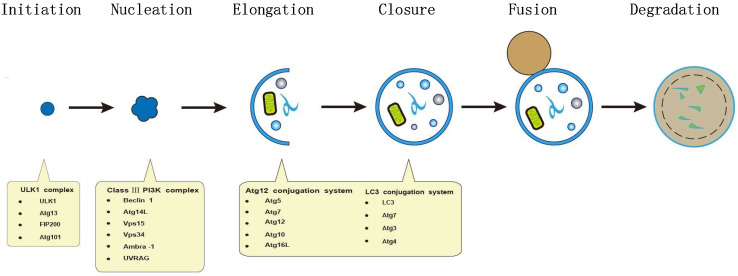
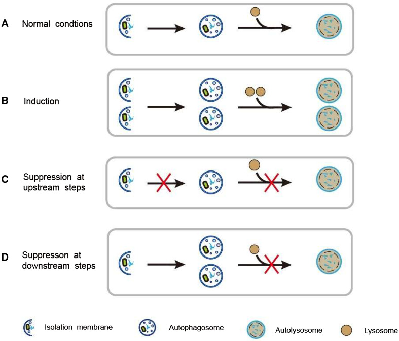
Axon degeneration is a pathophysiological process of axonal dying and breakdown, which is characterized by several morphological features including the accumulation of axoplasmic organelles, disassembly of microtubules, and fragmentation of the axonal cytoskeleton. Autophagy, a highly conserved lysosomal-degradation machinery responsible for the control of cellular protein quality, is widely believed to be essential for the maintenance of axonal homeostasis in neurons. In recent years, more and more evidence suggests that dysfunctional autophagy is associated with axonal degeneration in many neurodegenerative diseases. Here, we review the core machinery of autophagy in neuronal cells, and provide several major steps that interfere with autophagy flux in neurodegenerative conditions. Furthermore, this review highlights the potential role of neuronal autophagy in axon degeneration, and presents some possible molecular mechanisms by which dysfunctional autophagy leads to axon degeneration in pathological conditions.

Keywords: Autophagy; Axon degeneration; Mitophagy; Wallerian degeneration.


Conflict of interest statement

The authors declare no conflict of interest.

Figures

Fig. 1
Fig. 1
Cellular mechanism of autophagy. The autophagic pathway can be divided into separate stages, including initiation, nucleation, elongation, maturation, fusion, and degradation. The ULK1–Atg13–FIP200–Atg101 complex initiates autophagic process and facilitates the recruitment of other autophagy-related proteins to the site of autophagosome nucleation. Furthermore, the formation of autophagosomes requires the activity of the class III phosphatidylinositol 3-kinase (PI3K) consisting of Beclin 1, Atg14L, hVps15, and hVps34. Membranes originated from the ER, Golgi complex, or mitochondria compose the cup-like isolation membrane (also called phagophore). During the stage of elongation, LC3-II and the Atg12–Atg5–Atg16L complex locate on the membrane and promote phagophore expansion. In the meantime, phagophore engulfs cytoplasmic components, resulting in an autophagosome. Then, the double-membrane autophagosome merge with lysosome to form a single-membrane autolysosome. Finally, the components engulfed in autophagosome are degraded and released as small molecules
Fig. 2
Fig. 2
Dynamic process of autophagic flux under different conditions. a Under normal conditions, basal autophagy occurs. b When autophagy is induced, the formation of both autophagosomes and autolysosomes increases, and autophagic flux is enhanced. c When autophagy is suppressed at upstream steps, the formation of autophagosomes and autolysosomes decreases, and autophagic flux is blocked. d When autophagy is suppressed at downstream steps, only the number of autophagosomes increases, whereas the formation of autolysosomes is blocked. Thus, autophagic flux decreases
Fig. 3
Fig. 3
Proposed cellular pathways by which autophagy is implicated in Wallerian degeneration. Axonal injury can induce Wallerian degeneration, which is modulated by the opposing actions of pro-degenerative and pro-survival factors. It is supposed that autophagy may interact with pro-degenerative factors (SARM1, CRMP2 and calpain) and pro-survival factors (NMNATs, SIRT1 and SCG10) to modulate the process of Wallerian degeneration. However, the exact functions and underlying mechanisms of the critical molecules with respect to autophagy in Wallerian degeneration remain largely unknown

References

    1. Luo L, O’Leary DD. Axon retraction and degeneration in development and disease. Annu Rev Neurosci. 2005;28:127–156. doi: 10.1146/annurev.neuro.28.061604.135632. - DOI - PubMed
    1. Vargas ME, Barres BA. Why is Wallerian degeneration in the CNS so slow? Annu Rev Neurosci. 2007;30:153–179. doi: 10.1146/annurev.neuro.30.051606.094354. - DOI - PubMed
    1. Scheib J, Hoke A. Advances in peripheral nerve regeneration. Nat Rev Neurol. 2013;9:668–676. doi: 10.1038/nrneurol.2013.227. - DOI - PubMed
    1. Adalbert R, Morreale G, Paizs M, Conforti L, Walker SA, Roderick HL, Bootman MD, Siklos L, Coleman MP. Intra-axonal calcium changes after axotomy in wild-type and slow Wallerian degeneration axons. Neuroscience. 2012;225:44–54. doi: 10.1016/j.neuroscience.2012.08.056. - DOI - PubMed
    1. Raff MC, Whitmore AV, Finn JT. Axonal self-destruction and neurodegeneration. Science. 2002;296:868–871. doi: 10.1126/science.1068613. - DOI - PubMed

Publication types

LinkOut - more resources