. 2018 Jul;22(7):3489-3502.
doi: 10.1111/jcmm.13626. Epub 2018 Apr 19.

Endoplasmic reticulum stress induces spatial memory deficits by activating GSK-3

Affiliations

Endoplasmic reticulum stress induces spatial memory deficits by activating GSK-3

Li Lin et al. J Cell Mol Med. 2018 Jul.

Abstract

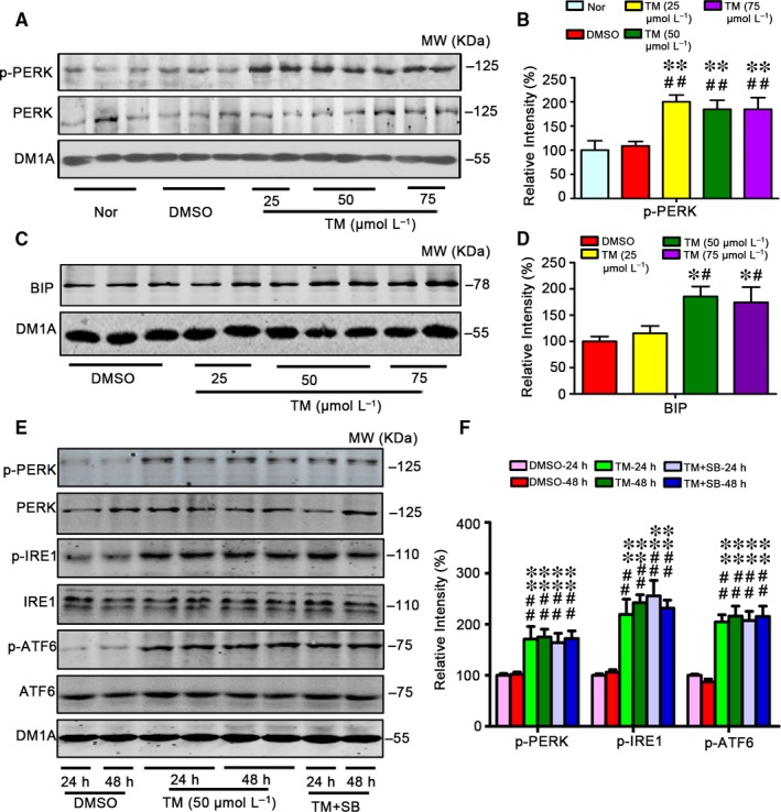
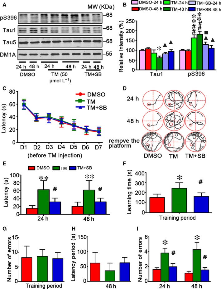
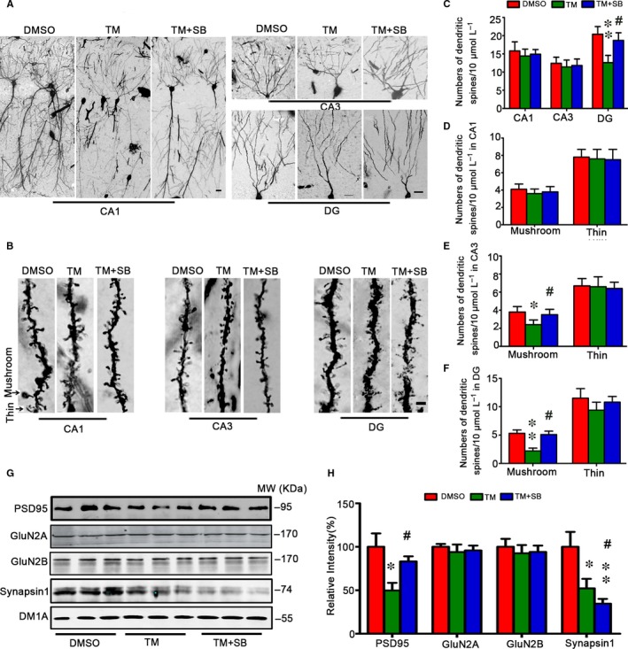
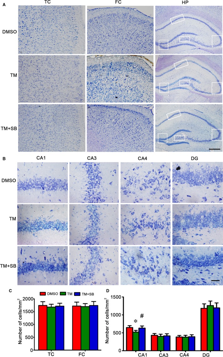
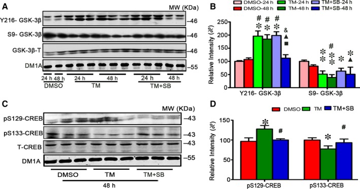
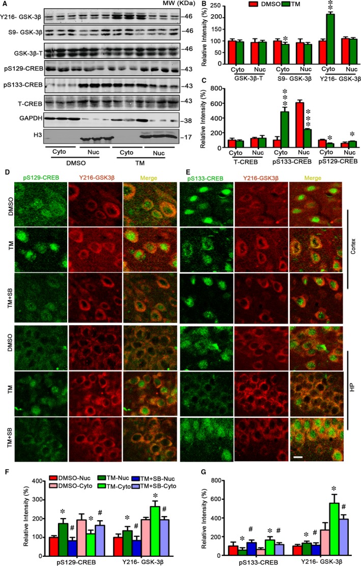
Endoplasmic reticulum (ER) stress is involved in Alzheimer's disease (AD), but the mechanism is not fully understood. Here, we injected tunicamycin (TM), a recognized ER stress inducer, into the brain ventricle of Sprague-Dawley (SD) rats to induce the unfolded protein response (UPR), demonstrated by the enhanced phosphorylation of pancreatic ER kinase (PERK), inositol-requiring enzyme-1 (IRE-1) and activating transcription factor-6 (ATF-6). We observed that UPR induced spatial memory deficits and impairments of synaptic plasticity in the rats. After TM treatment, GSK-3β was activated and phosphorylation of cAMP response element binding protein at Ser129 (pS129-CREB) was increased with an increased nuclear co-localization of pY126-GSK-3β and pS129-CREB. Simultaneous inhibition of GSK-3β by hippocampal infusion of SB216763 (SB) attenuated TM-induced UPR and spatial memory impairment with restoration of pS129-CREB and synaptic plasticity. We concluded that UPR induces AD-like spatial memory deficits with mechanisms involving GSK-3β/pS129-CREB pathway.

Keywords: Alzheimer's disease; cAMP response element binding protein; endoplasmic reticulum; glycogen synthase kinase-3; spatial memory deficits.


Figures

Figure 1
Figure 1
Tunicamycin induces UPR independent of GSK‐3 in rats. The male SD rats (4 m old) received ventricular infusion of 25, 50 or 75 μmol/L tunicamycin (TM, 10 μL) for 24 h (A,C), or infused with 50 μmol/L SB for 24 h and 48 h (E). The same volume of DMSO was infused as vehicle control, and the normal group (Nor) was killed without any treatment. The hippocampal extract was used for Western blotting (A,C,E) and quantitative analysis (B,D,F). The levels of the phosphorylated ER stress marker proteins as labelled except Bip were normalized against the total level, the latter and Bip were normalized against tubulin probed by DM1A. The data were expressed as means ± SD (n = 6). ** P < .01 vs Nor, ## P < .01 vs DMSO in B; * P < .05 vs DMSO, # P < .05 vs TM (25 μm) in D; ** P < .01 vs DMSO‐24 h, ## P < .01 vs DMSO‐48 h in F
Figure 2
Figure 2
SB attenuates tau hyperphosphorylation and memory deficits induced by tunicamycin in rats. The rats were randomly divided into 3 groups infused, respectively, through ventricle with 50 μmol/L TM or DMSO or TM plus SB (50 μmol/L). The rats were trained in Morris water maze (MWM) for 7 days before DMSO, TM and TM + SB injection (C). After 24 or 48 h, the brain extract from hippocampal regions (HP) was used to measure the alterations of tau proteins by Western blotting (A) and quantitative analysis (B). The levels of unphosphorylated tau at Tau1 epitope and the phosphorylation level of tau at Ser396 epitope as labelled on the blot were normalized against total tau probed by Tau5 which was normalized against DM1A (n = 6). SB could more obviously rescue the decreased Tau1 and the increased phosphorylation level of tau at Ser396 epitope after TM being injected for 48 h. Simultaneously, the MWM and step‐down avoidance tests were used to assess learning and memory capacities (D‐I). The rats had same cognitive levels during 7 days training before TM treatment (C), while injection of TM for 24 or 48 h induced memory deficits shown by the increased latency to find the hidden platform in MWM test (D,E). TM‐injected rats used more time to learn to protect themselves from the risk of electric shock in the training period during step‐down avoidance test measured at 24 and 48 h after the injection (F). TM‐injected rats showed no difference of the number of errors compared with the control group rats (G). TM‐injected rats showed no difference of latency period but increased number of errors in the detection period during step‐down avoidance test measured at 24 and 48 h after the injection. SB rescued TM‐induced memory deficits shown by the significantly decreased latency to find the hidden platform in MWM test (E). The data were expressed as mean ± SD (n = 10). * P < .05, ** P < .01 vs DMSO‐24 h; # P < .05, ## P < .01 vs DMSO 48 h; P < .05vs TM‐24 h, P < .05 vs TM‐48 h in B; * P < .05, ** P < .01 vs DMSO; # P < .05 vs TM in E‐I
Figure 3
Figure 3
Tunicamycin inhibits mushroom spine formation and expression of synaptic protein and attenuation by SB. The representative images of dendritic spines in rat hippocampal CA1, CA3 and DG at 48 h after TM injection (A,B). TM decreased numbers of mushroom‐type spines significantly in DG and CA3 subsets but not in CA1 of the TM‐treated group, and no significant change of thin‐spines was detected (B‐F). SB could reverse the above phenomenon. The levels of synapse‐associated proteins were measured by Western blotting and quantitative analysis, normalized against tubulin probed by DM1A (G,H). The data were expressed as mean ± SD (n = 3 for A and B, bar = 50 μm for A, bar = 2 μm for B; n = 6 for G). * P < .05, ** P < .05 vs DMSO, # P < .05 vs TM
Figure 4
Figure 4
Tunicamycin induces cell loss in hippocampal CA1 subset and attenuation by SB. The representative Nissl staining analysis shows temporal cortex (TC), frontal cortex (FC) and hippocampus (HP) after TM injection for 48 h (A). The neuronal numbers in hippocampal CA1, CA3, CA4 and DG were analysed (bar = 500 μm for TC, FC and HP; bar = 50 μm for CA1, CA3, CA4 and DG) (B,C). The data were expressed as mean ± SD (n = 6). * P < .05 vs DMSO, # P < .05 vs TM
Figure 5
Figure 5
Tunicamycin treatment affects GSK‐3 and CREB phosphorylation. After ventricular infusion of DMSO, TM at concentration of 50 μmol/L or TM plus SB (SB, 50 μmol/L) for 24 and 48 h, GSK‐3β levels of the hippocampal extract were measured by Western blotting (A) and quantitative analysis (B). SB treatment rescued the increased Y216‐GSK‐3β and decreased S9‐GSK‐3β induced by TM especially at 48 h. And for 48 h, SB treatment restored the levels of pSer133‐CREB and pSer129‐CREB measured by Western blotting (C) and quantitative analysis (D). The data were expressed as means ± SD (n = 6) * P < .05, ** P < .01 vs DMSO‐24 h, # P < .05 vs DMSO‐48 h, P < .05 vs TM‐24 h, P < .05 vs TM‐48 h, & P < .05 vs TM + SB‐24 h in B. * P < .05 vs DMSO, # P < .05 vs TM in D
Figure 6
Figure 6
Tunicamycin increases nuclear co‐localization of Y216‐GSK‐3β and pSer129‐CREB with reduced pSer133‐CREB. Rats were treated with TM or DMSO for 48 h and then the cytoplasmic and nuclear fractions of the hippocampus were analysed by Western blotting and quantitative analysis (A‐C). The representative immunofluorescence images show phosphorylated CREB probed by pSer133 and pSer129 (green), and phosphorylated GSK‐3β by Y216‐GSK‐3β (red) in cortex and hippocampus (bar = 50 μm) (D,E). SB treatment restored the nuclear internal and external metastasis of pSer133‐CREB and pSer129‐CREB. The co‐localization of pY216‐GSK‐3β with p‐S129‐CREB and p‐S133‐CREB in hippocampus were statistically measured. The data were expressed as means ± SD. The tendency of pY216‐GSK‐3β with p‐S129‐CREB and p‐S133‐CREB in cortex was similar that in hippocampus (F,G) and the statistical graph not be showed. (n = 6) * P < .05, ** P < .01, *** P < .001 vs DMSO in B and C. * P < .05 vs DMSO , # P < .05 vs TM in F and G

References

    1. Grundke‐Iqbal I, Iqbal K, Quinlan M, Tung YC, Zaidi MS, Wisniewski HM. Microtubule‐associated protein tau. A component of Alzheimer paired helical filaments. J Biol Chem. 1986;261:6084‐6089. - PubMed
    1. Huang HC, Jiang ZF. Accumulated amyloid‐beta peptide and hyperphosphorylated tau protein: relationship and links in Alzheimer's disease. J Alzheimers Dis. 2009;16:15‐27. - PubMed
    1. sKo¨pke E, Tung YC, Shaikh S, Alonso AC, Iqbal K, Grundke‐Iqbal I. Microtubule‐associated protein tau. Abnormal phosphorylation of a non‐paired helical filament pool in Alzheimer disease. J Biol Chem 1993;268:24374‐24384. - PubMed
    1. Alonso AC, Zaidi T, Grundke‐Iqbal I, Iqbal K. Role of abnormally phosphorylated tau in the breakdown of microtubules in Alzheimer disease. Proc Natl Acad Sci USA. 1994;91:5562‐5566. - PMC - PubMed
    1. Markesbery WR. Neuropathologic alterations in mild cognitive impairment: a review. J Alzheimers Dis. 2010;19:221‐228. - PMC - PubMed

Publication types

MeSH terms

LinkOut - more resources