. 2018 May 15;57(19):2814-2827.
doi: 10.1021/acs.biochem.8b00378. Epub 2018 May 3.

A High-Throughput Assay for Collagen Secretion Suggests an Unanticipated Role for Hsp90 in Collagen Production

Affiliations

A High-Throughput Assay for Collagen Secretion Suggests an Unanticipated Role for Hsp90 in Collagen Production

Madeline Y Wong et al. Biochemistry. .

Abstract

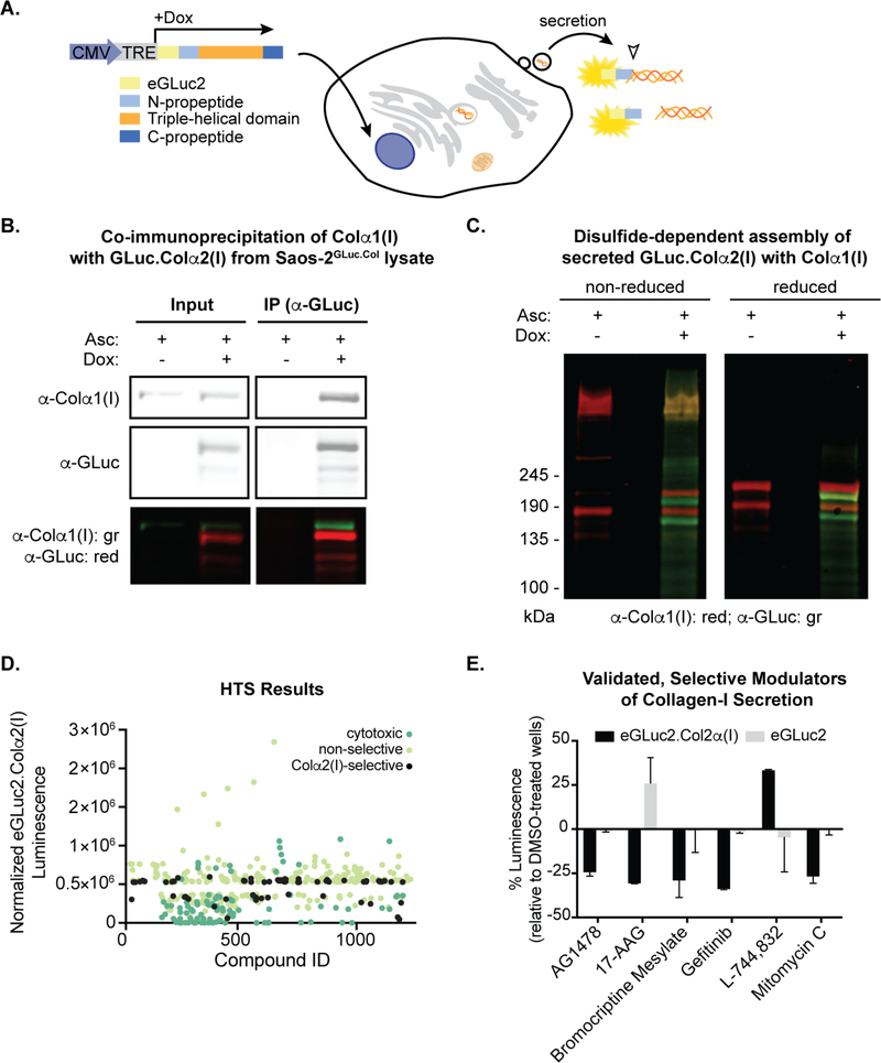
Collagen overproduction is a feature of fibrosis and cancer, while insufficient deposition of functional collagen molecules and/or the secretion of malformed collagen is common in genetic disorders like osteogenesis imperfecta. Collagen secretion is an appealing therapeutic target in these and other diseases, as secretion directly connects intracellular biosynthesis to collagen deposition and biological function in the extracellular matrix. However, small molecule and biological methods to tune collagen secretion are severely lacking. Their discovery could prove useful not only in the treatment of disease, but also in providing tools for better elucidating mechanisms of collagen biosynthesis. We developed a cell-based, high-throughput luminescent assay of collagen type I secretion and used it to screen for small molecules that selectively enhance or inhibit that process. Among several validated hits, the Hsp90 inhibitor 17-allylaminogeldanamycin (17-AAG) robustly decreases the secretion of collagen-I by our model cell line and by human primary cells. In these systems, 17-AAG and other pan-isoform Hsp90 inhibitors reduce collagen-I secretion post-translationally and are not global inhibitors of protein secretion. Surprisingly, the consequences of Hsp90 inhibitors cannot be attributed to inhibition of the endoplasmic reticulum's Hsp90 isoform, Grp94. Instead, collagen-I secretion likely depends on the activity of cytosolic Hsp90 chaperones, even though such chaperones cannot directly engage nascent collagen molecules. Our results highlight the value of a cell-based high-throughput screen for selective modulators of collagen secretion and suggest an unanticipated role for cytosolic Hsp90 in collagen secretion.


Conflict of interest statement

The authors declare that they have no conflicts of interest with the contents of this article.

Figures

Figure 1.
Figure 1.
High-throughput screening strategy and results. (A) Schematic of screen design. Addition of 1 μg/mL doxycycline (Dox) to the stable single-colony Saos-2GLuc.Col cell line induces expression of the eGLuc2.Colα2(I) fusion protein, which can be proteolytically processed post-secretion. Luminescence generated by the fusion protein may be assayed upon addition of the eGLuc2 substrate. (B) eGLuc2.Colα2(I) from Saos-2GLuc.Col lysate was immunoprecipitated with an α-GLuc antibody and analyzed by immunoblotting, revealing that eGLuc2.Colα2(I) associates intracellularly with Colα1(I). (C) Disulfide-dependent assembly of eGLuc2.Colα2(I) with Colα1(I). Collagen-I precipitated from conditioned media of Saos-2GLuc.Col cells was treated with iodoacetamide and heated in Laemmli buffer with or without DTT. Samples were separated by SDS-PAGE and analyzed by immunoblotting. (D) Plot of filtered screening results. (E) Validated small molecule modulators of collagen-I secretion.
Figure 2.
Figure 2.
Pan-Hsp90 inhibitors reduce endogenous collagen-I secretion. (A) Dose-response curve for the screening hit 17-AAG in Saos-2GLuc.Col cells. Cells were plated in 384-well plates and co-treated 5−6 h after plating with 1 μg/mL Dox and 17-AAG. After 23 h, plates were cooled at rt for 1–1.5 h, substrate was added, and luciferase activity was assayed. (B) Structures, names, and compound classes of tested pan-isoform Hsp90 inhibitors. (C) Western blot of conditioned media (top) and lysate (bottom) from primary dermal fibroblasts. Cells were plated and allowed to adhere overnight before receiving fresh media with 200 μM ascorbate and the following concentrations of Hsp90 inhibitors, as indicated: 17-AAG (250 nM), 17-DMAG (250 nM), EC 144 (100 nM), radicicol (250 nM), STA-9090 (300 nM). After 24 h of treatment, conditioned media and cells were harvested for immunoblotting. (D) Quantification of Western blot results. * = p < 0.05 (n = 3). (E) Western blot of conditioned media from a second healthy fibroblast line. * = p < 0.05; ** = p < 0.01 (n = 3).
Figure 3.
Figure 3.
Effects of Hsp90 inhibition on collagen-I secretion are post-translational. (A) FACS analysis of Annexin-V/Alexa Fluor 488 and propidium iodide staining to detect cell death. Cells were plated in triplicate for each condition and allowed to adhere overnight before being treated as in Fig. 2C. After 24 h, cells were trypsinized and labeled for FACS analysis. (B) qPCR analysis of collagen-I transcript levels from cells treated as in Fig. 2C. Average values are shown with the standard deviation for each condition (n = 3). (C) Measurement of collagen-I synthesis by [5-3H]proline pulse. Cells treated for 6 h with ascorbate and 17-AAG (250 nM) were pulsed with [5-3H]proline for 30 min prior to harvesting and analysis. Quantitation of newly synthesized collagen-I (mean ± standard deviation) is shown along with a representative gel image (n = 3).
Figure 4.
Figure 4.
Pan-Hsp90 inhibitors do not globally affect protein secretion. (A) Experimental workflow. Cells were plated and allowed to adhere overnight before being transduced with either GFP, transthyretin (TTR), or fibulin-3-expressing adenoviruses. The next day, transductions were split 1:2 and allowed to adhere overnight; cells were then treated with ascorbate and DMSO or 17-AAG (250 nM) for 24 h prior to harvesting. (B) Western blot of conditioned media from primary dermal fibroblasts transduced with FLAG-tagged transthyretin (TTR) or fibulin-3. (C) Immunoblot analysis of the secretion of endogenous fibronectin after treatment with 17-AAG. Cells were plated and treated as described in Fig. 2C. (D) Quantification of Western blot results. * = p < 0.05 (n = 3). (E) Phosphorimage of 35S-labeled secreted proteins. Cells were plated in triplicate for each condition and allowed to adhere overnight before being pre-treated with ascorbate and DMSO or 17-AAG (250 nM) for 2 h. Cells were then washed and cultured in radiolabeled media containing ascorbate, [35S]-Met/Cys, and DMSO / 17-AAG for 6 h. A representative radiograph is shown; the remaining biological replicates are presented in Fig. S5B.
Figure 5.
Figure 5.
Grp94 is dispensable for collagen-I secretion from primary fibroblasts. (A) Diagram of cellular Hsp90 isoforms along with the known collagen-I secretion pathway. Grp94, in the ER, is the only Hsp90 isoform capable of directly interacting with nascent collagen-I. (B) Western blot of lysate after lentiviral shRNA knockdown of Grp94. Cells were plated in triplicate for each construct and allowed to adhere overnight before being transduced with non-targeting control (nmc) or Grp94 shRNA lentiviruses. Media was changed the next day, and cells were left until 72 h post-transduction, when they received fresh media with ascorbate for 24 h prior to harvesting. (C) Immunoblots of corresponding media for the experiment described in (B). (D) Experimental workflow. Cells were plated in triplicate for each construct and allowed to adhere overnight before being transduced with non-targeting control or a Grp94 shRNA lentivirus. After 48 h, transductions were split 1:2 and allowed to adhere overnight. Cells subsequently received fresh media and were treated with ascorbate and DMSO or 17-AAG for 24 h prior to harvesting. (E) Immunoblots of conditioned media from cells co-treated with shRNA-containing lentivirus and DMSO or 17-AAG (n = 3).

References

    1. Brinckmann J (2005) Collagens at a glance, Top. Curr. Chem. 247, 1–6.
    1. Ricard-Blum S (2011) The collagen family, CSHL Persp. Biol. 3, a004978. - PMC - PubMed
    1. An B, Lin YS, and Brodsky B (2016) Collagen interactions: Drug design and delivery, Adv. Drug Deliv.Rev. 97, 69–84. - PubMed
    1. Heino J (2007) The collagen family members as cell adhesion proteins, Bioessays 29, 1001–1010. - PubMed
    1. Ishikawa Y, and Bachinger HP (2013) A molecular ensemble in the rER for procollagen maturation, Biochim. Biophys. Acta 1833, 2479–2491. - PubMed

Publication types

MeSH terms

LinkOut - more resources