. 2018 Apr 20;12(4):e0006446.
doi: 10.1371/journal.pntd.0006446. eCollection 2018 Apr.

SmSP2: A serine protease secreted by the blood fluke pathogen Schistosoma mansoni with anti-hemostatic properties

Affiliations

SmSP2: A serine protease secreted by the blood fluke pathogen Schistosoma mansoni with anti-hemostatic properties

Adrian Leontovyč et al. PLoS Negl Trop Dis. .

Abstract

Background: Serine proteases are important virulence factors for many pathogens. Recently, we discovered a group of trypsin-like serine proteases with domain organization unique to flatworm parasites and containing a thrombospondin type 1 repeat (TSR-1). These proteases are recognized as antigens during host infection and may prove useful as anthelminthic vaccines, however their molecular characteristics are under-studied. Here, we characterize the structural and proteolytic attributes of serine protease 2 (SmSP2) from Schistosoma mansoni, one of the major species responsible for the tropical infectious disease, schistosomiasis.

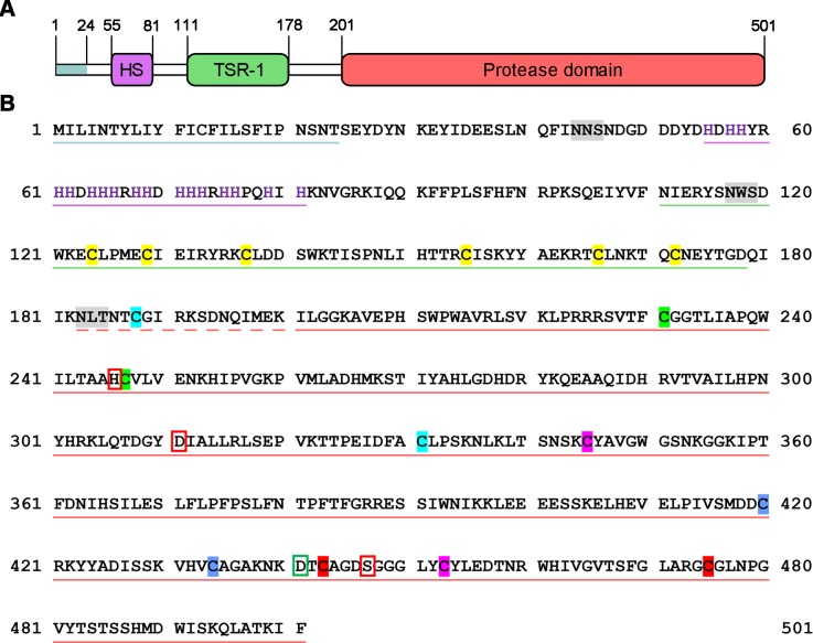
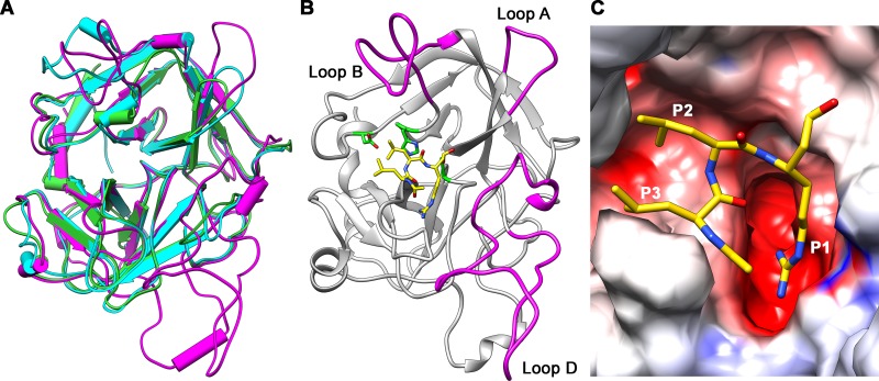
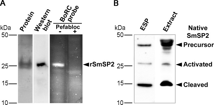
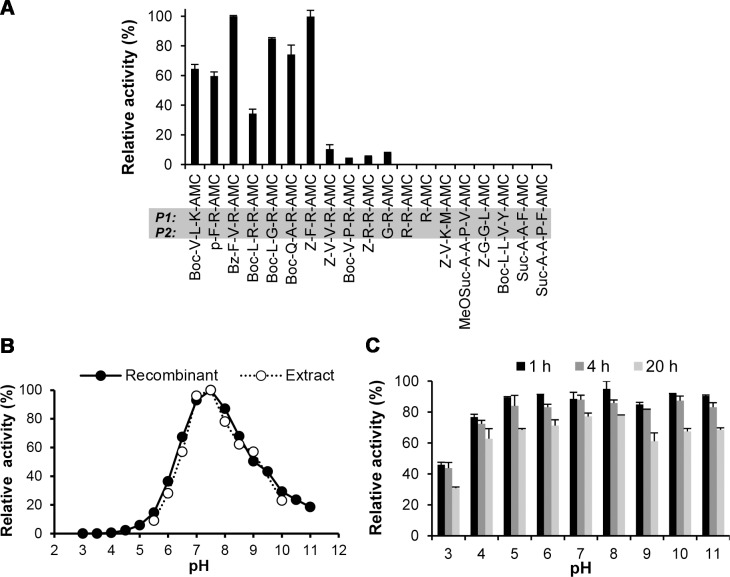
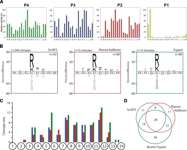
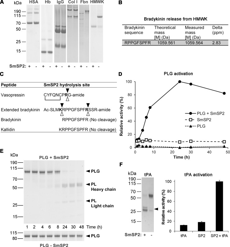
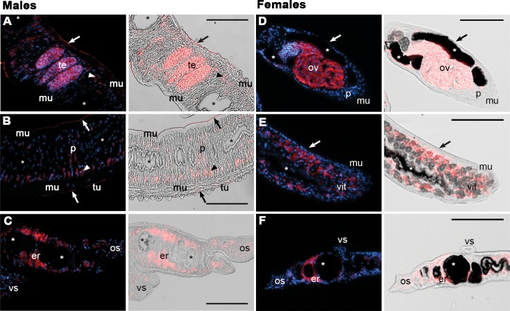
Methodology/principal findings: SmSP2 comprises three domains: a histidine stretch, TSR-1 and a serine protease domain. The cleavage specificity of recombinant SmSP2 was determined using positional scanning and multiplex combinatorial libraries and the determinants of specificity were identified with 3D homology models, demonstrating a trypsin-like endopeptidase mode of action. SmSP2 displayed restricted proteolysis on protein substrates. It activated tissue plasminogen activator and plasminogen as key components of the fibrinolytic system, and released the vasoregulatory peptide, kinin, from kininogen. SmSP2 was detected in the surface tegument, esophageal glands and reproductive organs of the adult parasite by immunofluorescence microscopy, and in the excretory/secretory products by immunoblotting.

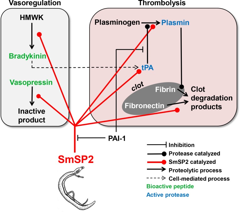
Conclusions/significance: The data suggest that SmSP2 is secreted, functions at the host-parasite interface and contributes to the survival of the parasite by manipulating host vasodilatation and fibrinolysis. SmSP2 may be, therefore, a potential target for anti-schistosomal therapy.


Conflict of interest statement

The authors have declared that no competing interests exist.

Figures

Fig 1
Fig 1. Domain organization and amino acid sequence of SmSP2.
(A) Schematic diagram of the domain layout. The N-terminal signal peptide, “His stretch’ (HS), thrombospondin type 1 repeat (TSR-1) and S1 family protease domains are depicted in blue, purple, green, and red, respectively. Amino acid residue numbers are indicated. (B) The amino acid sequence of SmSP2 with the various domains color-coded by underlining as in (A). Predicted N-glycosylation sites are highlighted in grey, and His residues in the His stretch are in purple. The catalytic residues, His246, Asp311 and Ser447 are red-boxed; and Asp441 in the S1 subsite that accounts for trypsin-like activity is green-boxed. Cys residues of the protease domain that are predicted to form a disulfide are indicated by the same color; Cys residues of the TSR-1 domain are colored yellow. The propeptide is underlined with dashed red line.
Fig 2
Fig 2. Homology model of the SmSP2 protease domain.
The model was built using as a template the X-ray structures of bovine trypsin (PDB 1JRT) and human MASP-1 (PDB 3GOV). (A) A superposition of the SmSP2 model (magenta), the bovine trypsin (green) and MASP-1 (cyan) crystal structures in a cylinder representation. (B) A view from the top on the SmSP2 active site with covalently bound substrate-like inhibitor leupeptin (N-acetyl-L-leucyl-L-leucyl-L-argininal). Carbon atoms of leupeptin are yellow; heteroatoms have the standard color coding (N, blue; O, red). SmSP2 catalytic residues are green. The active site is partially blocked by loops A, B and D (magenta) that are formed by insertions in SmSP2 sequence compared to trypsin sequence. (C) A surface representation of the SmSP2 active site colored by electrostatic potential (at a scale from -10 kT/e (red) to +10 kT/e (blue)). Inhibitor leupeptin is colored as in (B).
Fig 3
Fig 3. Preparation of recombinant SmSP2 and identification of native SmSP2.
(A) The recombinant protease domain of SmSP2 (rSmSP2) expressed in P. pastoris was resolved by SDS-PAGE and protein-stained or visualized by polyclonal anti-rSmSP2 IgG. For in-gel activity-based labeling, rSmSP2 was incubated with the fluorescent active site probe, BoRC, resolved by SDS-PAGE and visualized using a fluorescence scanner. The competitive labeling was performed with the serine protease inhibitor, Pefabloc SC. (B) Protein extracts of S. mansoni adult worms and their ESP were resolved by SDS-PAGE and visualized by the anti-rSmSP2 IgG.
Fig 4
Fig 4. Substrate specificity and pH profile of SmSP2.
(A) Activity of rSmSP2 was probed using a panel of peptidyl fluorogenic substrates used to assay trypsin-like and chymotrypsin/elastase-like serine proteases. Substrate hydrolysis was measured in a kinetic assay at pH 8.0. The mean values ± S.D. of three replicates are normalized to the maximum value. Amino acid residues at P1 and P2 positions are highlighted by the grey bar. (B) The pH profiles of rSmSP2 and native SmSP2 activity in extracts of adult worms. Activity was measured in a kinetic assay using the fluorogenic substrate P-F-R-AMC. The native activity (sensitive to the serine protease inhibitor Pefabloc SC) was measured in the presence of 10 μM E-64 and 1 mM EDTA to prevent undesired proteolysis of the substrate by cysteine proteases and metalloproteases, respectively. The mean values of three replicates, expressed as a percentage normalized to the highest value, are shown (standard deviation values are within 5% of the mean). (C) The pH stability of rSmSP2. Activity of rSmSP2 was measured at pH 8.0 in a kinetic assay as in (B) after incubation of the enzyme at pH 3 to 11 for different times. The mean values of three replicates, expressed as a percentage normalized to activity of non-incubated rSmSP2, are shown.
Fig 5
Fig 5. Substrate specificity of rSmSP2.
(A) The P1 to P4 specificity of rSmSP2 was determined by a positional scanning-synthetic combinatorial library. The X-axis indicates 20 amino acids held constant at each position (n is norleucine). The Y-axis represents activity related to the most preferred amino acid (100%). (B) The P4 to P4′ specificity profiles of rSmSP2, human plasma kallikrein, and bovine trypsin were obtained using a multiplex combinatorial library (MSP-MS). The iceLogo substrate profiles were generated from the pattern of cleavage events after incubation with the 14-mer library. Amino acids that are most frequently found at each position are shown above the horizontal line, whereas amino acids that less frequently observed are below. (C) Spatial distribution of cleavage sites within the 14-mer peptide scaffold. (D) The Venn diagram shows the number of unique and shared cleavage sites.
Fig 6
Fig 6. Processing of host-derived proteins and peptides by rSmSP2.
(A) Human serum albumin (HSA), hemoglobin (Hb), immunoglobulin G (IgG), collagen type I (Col I), fibronectin (Fbn) and high molecular weight kininogen (HMWK) were incubated for 16 h at pH 8.0 in the presence (+) or absence (-) of rSmSP2. The reaction mixtures were subjected to SDS-PAGE and protein stained. (B) HMWK was incubated with rSmSP2 and the reaction mixture was subjected to LC-MS/MS analysis to identify bradykinin peptide released from HMWK. (C) Peptide hormones were incubated with rSmSP2 or with live adults maintained in culture and the cleavage positions (full triangles for rSmSP2, open triangles for adult schistosomes) were identified by mass spectrometry. Residues at the P1 position are in bold and the disulfide connectivity of vasopressin is indicated. (D) Human plasminogen (PLG) was incubated in the presence or absence of rSmSP2 and the reaction mixture was analyzed at different time points. Plasmin proteolytic activity generated during plasminogen processing by rSmSP2 was determined in a kinetic assay with Boc-V-L-K-AMC. Mean values of triplicates are expressed relative to the maximum value (100%). The S.D. values of three replicates are within 10% of the mean. All experiments were performed at least twice with similar results. (E) The processed forms were resolved by SDS-PAGE and visualized by protein staining. The positions for PLG, and plasmin (PL) heavy and light chains are indicated. (F) Human tissue plasminogen activator (tPA) was incubated for 16 h at pH 8.0 in the presence/absence of rSmSP2 and analyzed by SDS-PAGE with protein staining; proteolytic activity generated during tPA processing was monitored in a kinetic assay using Z-G-G-R-AMC. Mean values ± s.d. of triplicates are expressed relative to the maximum value (100%). Two chain tPA is indicated with an arrow.
Fig 7
Fig 7. SmSP2 immunolocalization in sections of adult S. mansoni.
Semi-thin sections of adult S. mansoni males (A to C) and females (D to F) were probed with an anti-SmSP2 IgG followed by reaction with an anti-rabbit Alexa 647-labeled secondary antibody (red). DAPI was used to label nuclear DNA (blue). The left columns show merged fluorescent channels; in the right columns, the signal is merged with differential interference contrast. A strong SmSP2 signal (red) was detected in both sexes in the parenchyma (p) and the esophageal region (er). A faint signal was noted in the ventral (vs) and oral suckers (os). No signal was detected in the gut (asterisks), muscle (mu) or tegumental tubercles (tu). In males, SmSP2 signal also appears in the tegument (arrowhead) in the tegumental membrane surface (arrow) and in the testes (te). In females, the signal is noted in the ovaries (ov) and vitellaria (vit). The scale bar represents 100 μm. A and D, reproductive organs; B and E, tegumental cells; C and F, head.
Fig 8
Fig 8. Model of how SmSP2 may interfere with the host’s hemostatic system.
SmSP2, secreted from adult schistosomes or localized at the surface, stimulates the degradation of blood clots (thrombolysis panel) by (i) activation of two critical components of the fibrinolytic system, tissue plasminogen activator (tPA) and plasminogen, and (ii) direct degradation of the blood-clot component, fibronectin. SmSP2 modulates vascular tone (vasoregulation panel) by processing bioactive peptide hormones. (i) It releases the vasodilatory bradykinin from kininogen (HMWK) and (ii) degrades the vasoconstrictory peptide, vasopressin. Bradykinin may stimulate the release of tPA from vascular endothelial cells (dashed line) which would promote fibrinolysis. SmSP2 may be regulated by plasminogen activator inhibitor-1 (PAI-1) that inhibits SmSP2 (Table 1).

References

    1. Steinmann P, Keiser J, Bos R, Tanner M, Utzinger J. Schistosomiasis and water resources development: systematic review, meta-analysis, and estimates of people at risk. Lancet Infect Dis. 2006;6(7):411–25. S1473-3099(06)70521-7 [pii]; doi: 10.1016/S1473-3099(06)70521-7 - DOI - PubMed
    1. King CH. Parasites and poverty: the case of schistosomiasis. Acta Trop. 2010;113(2):95–104. doi: 10.1016/j.actatropica.2009.11.012 ; PubMed Central PMCID: PMCPMC2812649. - DOI - PMC - PubMed
    1. Gryseels B, Polman K, Clerinx J, Kestens L. Human schistosomiasis. Lancet. 2006;368(9541):1106–18. doi: 10.1016/S0140-6736(06)69440-3 - DOI - PubMed
    1. Burke ML, Jones MK, Gobert GN, Li YS, Ellis MK, McManus DP. Immunopathogenesis of human schistosomiasis. Parasite Immunol. 2009;31(4):163–76. PIM1098 [pii]; doi: 10.1111/j.1365-3024.2009.01098.x - DOI - PubMed
    1. Caffrey CR. Chemotherapy of schistosomiasis: present and future. Curr Opin Chem Biol. 2007;11(4):433–9. doi: 10.1016/j.cbpa.2007.05.031 - DOI - PubMed

Publication types

MeSH terms