. 2018 Apr 20;13(4):e0192709.
doi: 10.1371/journal.pone.0192709. eCollection 2018.

Osteopontin plays a pivotal role in increasing severity of respiratory syncytial virus infection

Affiliations

Osteopontin plays a pivotal role in increasing severity of respiratory syncytial virus infection

Viviana Sampayo-Escobar et al. PLoS One. .

Abstract

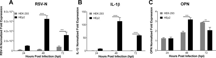
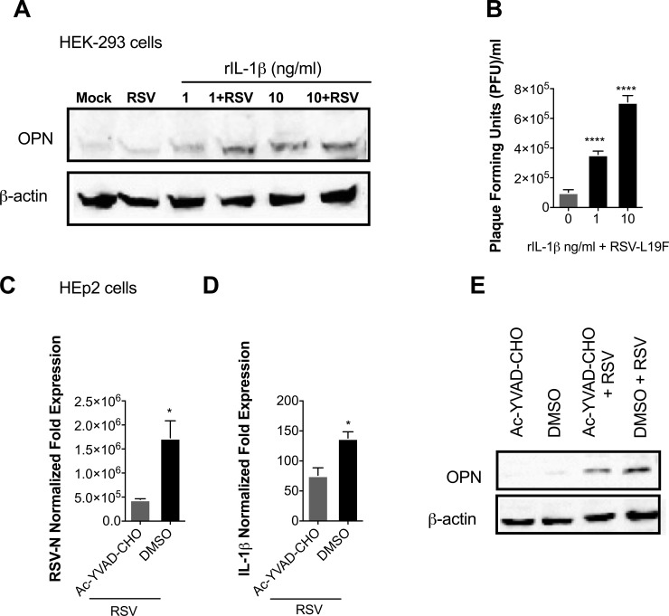
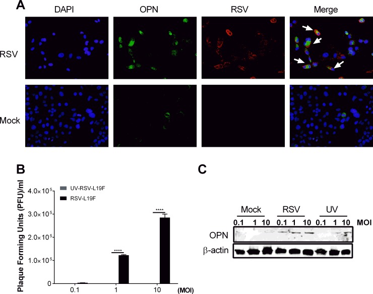
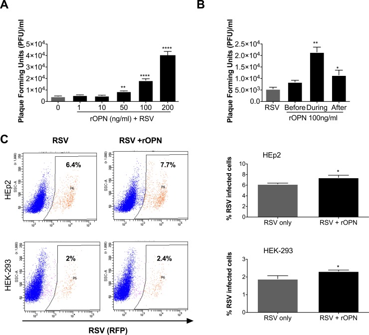
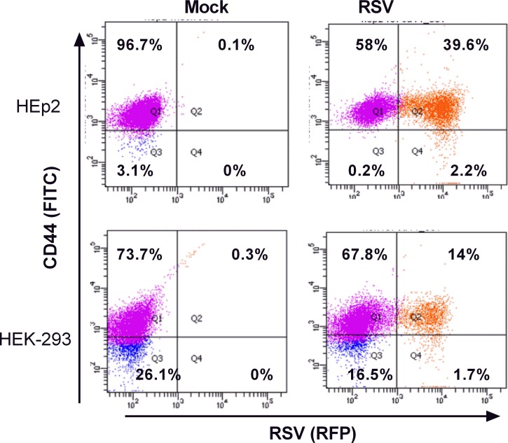
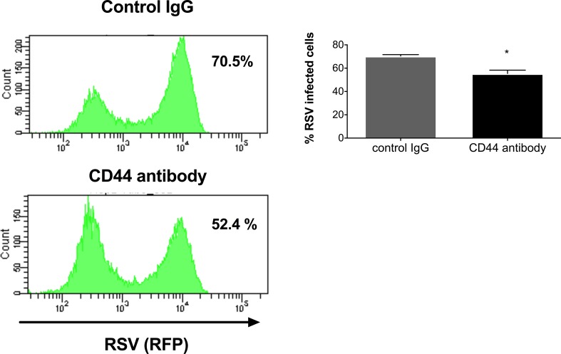
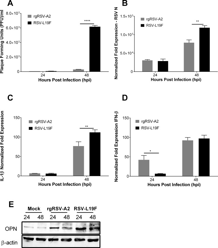
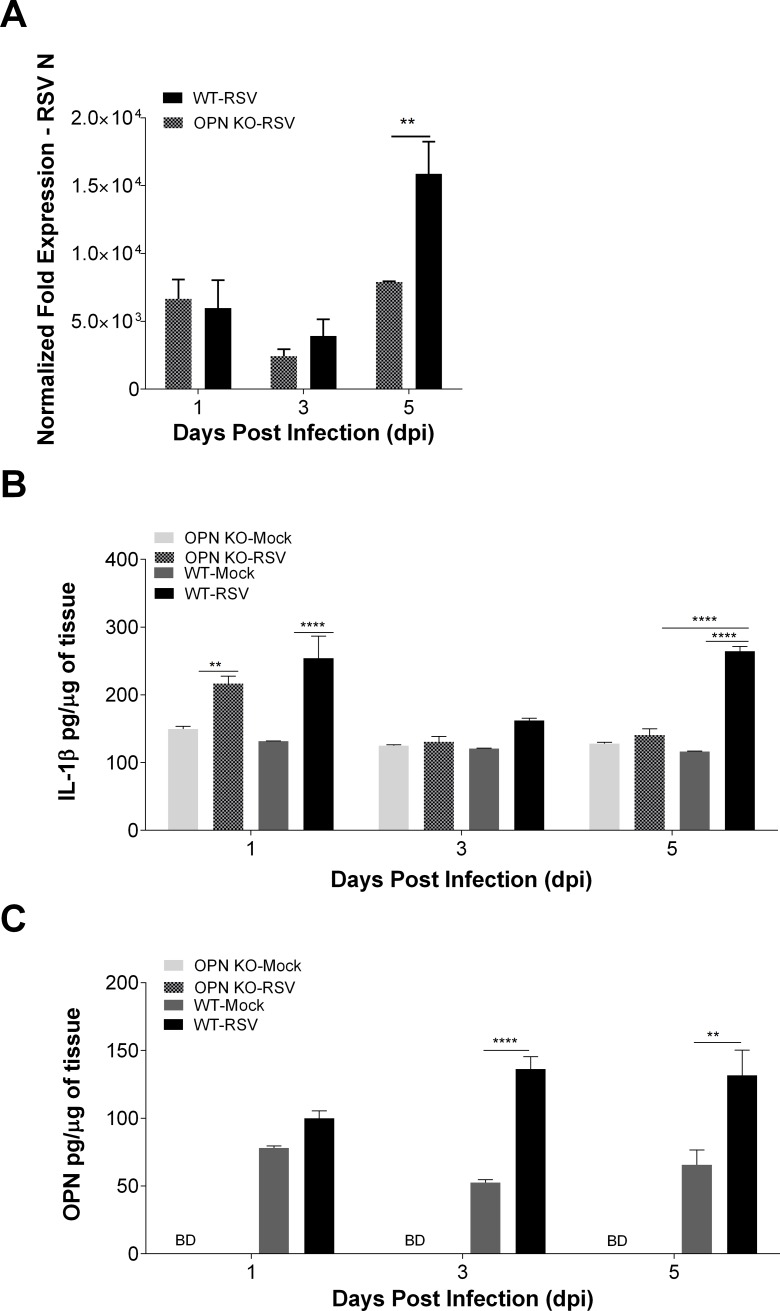
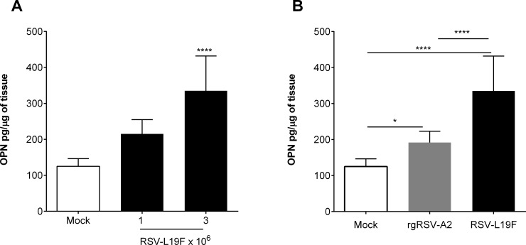
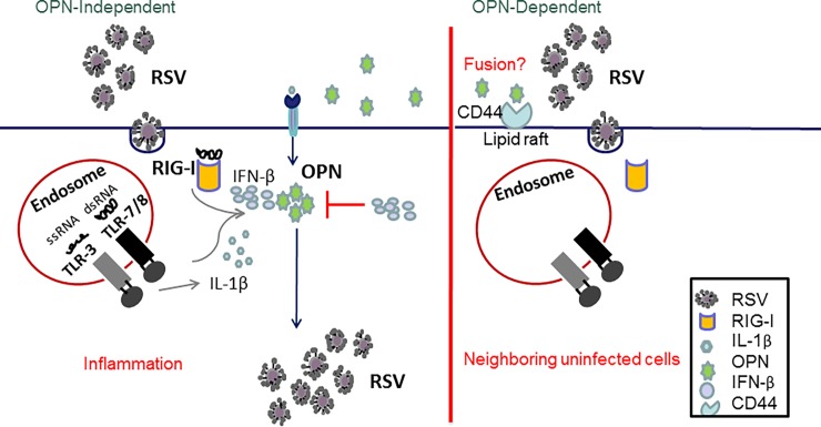
The molecular mechanisms underlying susceptibility to severe respiratory syncytial virus (RSV) infection remain poorly understood. Herein, we report on the role of osteopontin (OPN) in regulation of RSV infection in human epithelial cells and how interleukin-1 beta (IL-1β), a cytokine secreted soon after RSV infection, when persistently expressed can induce OPN expression leading to increased viral infection. We first compared OPN expression in two human epithelial cell lines: HEK-293 and HEp-2. In contrast to HEp-2, HEK-293 expresses low levels of pro-caspase-1 resulting in decreased IL-1β expression in response to RSV infection. We found a correlation between low IL-1β levels and a delay in induction of OPN expression in RSV-infected HEK-293 cells compared to HEp-2. This phenomenon could partially explain the high susceptibility of HEp-2 cells to RSV infection versus the moderate susceptibility of HEK-293 cells. Also, HEK-293 cells expressing low levels of pro-caspase-1 exhibit decreased IL-1β expression and delayed OPN expression in response to RSV infection. HEK-293 cells incubated with human rIL-1β showed a dose-dependent increase in OPN expression upon RSV infection. Also, incubation with rOPN increased RSV viral load. Moreover, HEp-2 cells or mice infected with a mucogenic RSV strain RSV-L19F showed elevated levels of OPN in contrast to mice infected with the laboratory RSV strain rA2. This correlated with elevated levels of OPN following infection with RSV-L19F compared to rA2. Together, these results demonstrate that increased OPN expression is regulated in part by IL-1β, and the interplay between IL-1β and OPN signaling may play a pivotal role in the spread of RSV infection.


Conflict of interest statement

Competing Interests: The authors have declared that no competing interests exist.

Figures

Fig 1
Fig 1. IL-1β and OPN mRNA expression in RSV infected cells.
HEp-2 and HEK-293 cells were mock infected or infected with 1 MOI of RSV-L19F. RNA was isolated at 24, 48 and 72 hpi. (A-C) Expression levels of RSV-N, IL-1β and OPN were determined by qPCR. Results are presented as fold-change in expression of RSV-N, IL-1β or OPN mRNA normalized to the control (HPRT). qPCR data are represented as means ±SEM. Experiments were performed in triplicate. ** p < 0.01, *** p < 0.001**** p < 0.0001.
Fig 2
Fig 2. RSV induced IL-1β regulates OPN expression during RSV infection.
(A-B) HEK-293 cells were mock-infected or infected with 1 MOI of RSV-L19F. 2 hours after the infection cells were treated with increasing concentrations of human rIL-1β (0, 1 or 10 ng/ml). (A) Protein was harvested 24 hpi for western blots. 25μg of protein lysate was loaded in each lane. OPN is seen at 55 kDa and β-actin (loading control) at 42 kDa. (B) Plaque viral titers were obtained from the supernatants collected 24 hpi. (C-E) HEp-2 cells were pre-treated two hours prior to RSV-L19F infection with 10 μM of Ac-YVAD-CHO (caspase-I) inhibitor. Cells were infected with 1 MOI of RSV-L19F and infectious media was replaced with fresh media containing 10 μM of Ac-YVAD-CHO. RNA and protein were collected 24 hpi. (C and D) Expression levels of IL-1β and RSV-N were determined by qPCR. Results are presented as fold-change in expression of IL-1β or RSV-N mRNA normalized to the control (HPRT). (E) 25μg of protein lysate was loaded in each lane. OPN is seen at 55 kDa and β-actin (loading control) at 42 kDa. qPCR data are represented as means ±SEM. Experiments were performed in triplicate. **** p < 0.0001.
Fig 3
Fig 3. RSV infection induces OPN expression in a dose dependent manner.
HEp-2 cells were mock-infected or infected with RSV-L19F (1 MOI). (A) Images of Hep-2 cells stained with polyclonal antibody against RSV and OPN at 48 hpi. Representative images of RSV positive (red) cells, OPN positive (green) cells, and DAPI (blue) nuclear staining at 200X magnification. (B) RSV titer by plaque assay of supernatants for increasing doses (0.1, 1 and 10 MOI) of UV-inactivated and native RSV-L19F at 48 hpi. (C) OPN protein (55 kDa) expression in HEp-2 cells at 48 hpi after mock, RSV and UV-inactivated RSV treatment in the indicated doses analyzed by western blots. β-actin (loading control) at 42 kDa was probed as the loading control.
Fig 4
Fig 4. Human rOPN increases RSV titers in a dose-dependent manner.
(A) HEK-293 cells were treated with increasing concentrations of human recombinant OPN (rOPN) (0 to 200 ng/ml) four hours before infection. Cells were infected with 0.1 MOI of RSV-L19F and infectious media was replaced with fresh media containing the appropriate concentration of rOPN. Supernatants were collected 24 hpi and viral titers were determined by plaque assay. (B) HEK-293 cells were treated with 100 ng/ml at different time points: before, during or immediately after the infection. Supernatants were collected 24 hpi and viral titers were determined by plaque assay. Result of a representative experiment is shown. * p < 0.05, ** p < 0.01, **** p < 0.0001. (C) Hep-2 or HEK-293 cells were infected with 0.1 MOI of RSV-KL19F or RSV-KL19F in the presence of 100 ng/ml of rOPN. After 24 hours, the infected HEp-2 or HEK-293 cells were gated for RFP-expression (RSV + cells).
Fig 5
Fig 5. CD44 expression mediates RSV infection.
HEp-2 or HEK-293 cells were infected with 0.5 MOI of RSV-KL19F (red). After 24 hours, the cells were stained with FITC mouse anti-human CD44 antibody (green) and gated for RSV expression (red). (Top panel) Flow cytometry analysis of HEp-2 cells infected with RSV-KL19F. (Bottom panel) Flow cytometry analysis of HEK-293 cells infected with RSV-KL19F. Result of a representative experiment is shown. Experiments were performed in triplicate.
Fig 6
Fig 6. Neutralization of CD44 receptor reduces the number of RSV positive cells.
Hep-2 cells were pre-treated with 10 μg of broad spectrum rat-anti human CD44 antibody (clone A020) or normal rat IgG (control). After the pre-treatment, cells were infected with RSV-KL19F (0.5 MOI) and infectious media was replaced with fresh growth media. After 24 hours, the percentage of RSV positive cells was determined by flow cytometry. Result of a representative experiment is shown. Experiments were performed in triplicate.
Fig 7
Fig 7. OPN is a marker of high RSV loads in HEp-2 cells.
HEp-2 cells were mock-infected or infected with 1 MOI of RSV-L19F or rgRSV-A2. RNA, supernatants and protein were isolated 24 and 48 hpi. (A) Plaque titers were obtained from supernatants of HEp-2 cells infected with RSV-L19F or rgRSV-A2. (B—D) Expression of RSV-N, IL-1β and IFN-β were determined by qPCR. Results are presented as fold-change in expression of RSV-N, IL-1β or IFN-β mRNA normalized to the control (HPRT). (E) Western blot analysis of OPN expression. 25μg of protein lysate was loaded in each lane. OPN is seen at 55 kDa and β-actin (loading control) at 42 kDa. Experiments were performed in triplicate. * p < 0.05, ** p < 0.01, **** p < 0.0001.
Fig 8
Fig 8. RSV replication is diminished in mice lacking OPN (OPN KO).
C57BL/6 and OPN KO mice were mock-infected or infected with 3 x 10⁶ RSV-L19F. Lungs were collected 1, 3 and 5 dpi for protein and RNA extraction. (A) RSV-N mRNA expression levels were determined by qPCR and results represented as fold-change in expression of RSV-N mRNA normalized to the control (HPRT). (B and C) Levels of OPN and IL-1β in lung homogenates were determined by ELISA. BD: Below detection limits. Lung homogenates were obtained from individual mice and not pooled (n>4 per group). ** p < 0.01, **** p < 0.0001.
Fig 9
Fig 9. OPN expression is a marker of high RSV loads in vivo.
(A) C57BL/6 mice were mock-infected or infected with 1 or 3 x 10⁶ RSV-L19F. (B) C57BL/6 mice were mock-infected or infected with 3 x 10⁶ RSV-L19F or rgRSV-A2. (A-B) Lungs were collected 3 dpi for protein isolation and levels of OPN in lung homogenates were determined by ELISA. Lung homogenates were obtained from individual mice and not pooled (n>4 per group). * p < 0.05, ** p < 0.01, **** p < 0.0001.
Fig 10
Fig 10. Model for the role of OPN during RSV infection.
(Left panel) OPN-independent model. During RSV infection, the virus is recognized by the host cells through PRR (TLR-3, RIG-I, TLR-7); recognition of the virus leads to activation of innate immune response which leads to release of anti-viral and pro-inflammatory cytokines like IFN-β and IL-1β. Expression of IL-1β leads to up-regulation of OPN whereas expression of IFN-β down-regulates OPN expression. (Right panel) OPN-dependent model. Fusion of RSV is accelerated in the presence of OPN and its receptor (CD44), thus leading to increased number of infected cells which results in increased viral loads.

References

    1. Piedimonte G, Perez MK. Alternative mechanisms for respiratory syncytial virus (RSV) infection and persistence: could RSV be transmitted through the placenta and persist into developing fetal lungs? Current Opinion in Pharmacology. 2014;16(0):82–8. 10.1016/j.coph.2014.03.008. - DOI - PMC - PubMed
    1. Hall CB, Walsh EE, Long CE, Schnabel KC. Immunity to and Frequency of Reinfection with Respiratory Syncytial Virus. Journal of Infectious Diseases. 1991;163(4):693–8. 10.1093/infdis/163.4.693 - DOI - PubMed
    1. Scott PD, Ochola R, Ngama M, Okiro EA, James Nokes D, Medley GF, et al. Molecular Analysis of Respiratory Syncytial Virus Reinfections in Infants from Coastal Kenya. Journal of Infectious Diseases. 2006;193(1):59–67. 10.1086/498246 - DOI - PMC - PubMed
    1. Borchers AT, Chang C, Gershwin ME, Gershwin LJ. Respiratory Syncytial Virus—A Comprehensive Review. Clinical Reviews in Allergy & Immunology. 2013;45(3):331–79. 10.1007/s12016-013-8368-9 - DOI - PMC - PubMed
    1. Collins PL, Graham BS. Viral and Host Factors in Human Respiratory Syncytial Virus Pathogenesis. Journal of Virology. 2008;82(5):2040–55. 10.1128/JVI.01625-07 - DOI - PMC - PubMed

Publication types

MeSH terms