Review
. 2018 Apr 26:36:309-338.
doi: 10.1146/annurev-immunol-042617-053245.

Complement and the Regulation of T Cell Responses

Affiliations
Review

Complement and the Regulation of T Cell Responses

Erin E West et al. Annu Rev Immunol. .

Abstract

The complement system is an evolutionarily ancient key component of innate immunity required for the detection and removal of invading pathogens. It was discovered more than 100 years ago and was originally defined as a liver-derived, blood-circulating sentinel system that classically mediates the opsonization and lytic killing of dangerous microbes and the initiation of the general inflammatory reaction. More recently, complement has also emerged as a critical player in adaptive immunity via its ability to instruct both B and T cell responses. In particular, work on the impact of complement on T cell responses led to the surprising discoveries that the complement system also functions within cells and is involved in regulating basic cellular processes, predominantly those of metabolic nature. Here, we review current knowledge about complement's role in T cell biology, with a focus on the novel intracellular and noncanonical activities of this ancient system.

Keywords: CD46; Th1 response; autoimmunity; complosome; infection; metabolism.


Figures

Figure 1
Figure 1
Activation and functions of serum-circulating complement. Liver-derived, systemically circulating complement can be activated through the classical pathway, the lectin pathway, and the alternative pathway. Through the formation of classical (C4b2b) and alternative (C3bBb) C3 convertases, these pathways lead to the generation of C3a and C3b. Upon subsequent generation of C5 convertase (C4bC2aC3b for the classical and lectin pathways and C3bBbC3b for the alternative pathway), C5b and C5a are produced, with surface-bound C5b initiating the formation and insertion of the MAC on pathogens (or other target membranes), and the generated anaphylatoxins act on innate immune cells to foster an inflammatory reaction. Complement activation is regulated by multiple inhibitory proteins (denoted in ‘red’ text blocks), such as CD46, CD55, CR1, Factor H, and C4BP, which aid in the inactivation of deposited C3b and C4b (CD46, Factor H, C4BP, and CR1) and/or disassemble the convertases (CR1 and CD55), whereas CD59 and vitronectin inhibit MAC formation. Abbreviations: C4BP, C4b-binding protein; CD46, membrane cofactor protein (MCP); CTSL, cathepsin L; FB, Factor B; FD, Factor D; MAC, membrane attack complex; MASP2, mannose-binding lectin serine protease 2; PAMPs, pathogen-associated molecular patterns.
Figure 2
Figure 2
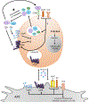
Non-canonical functions of immune cell-derived local complement. Local complement activation is triggered when activating signals (TCR stimulation on T cells and TLR activation on APCs) initiate the generation and secretion of C3, C5, Factor B, and Factor D, leading to C3 and C5 convertase formation in the extracellular space and on the cell surface and ultimately the generation of the complement activation fragments C3a, C3b, C5b, and C5a (C3 and C5 can also be cleaved by proteases in the extracellular space). TCR stimulation also induces the shuttling of C3aR from the lysosome to the cell surface, allowing these complement fragments to bind to their respective receptors on the cell surface and induce cellular responses (in conjunction with CD46 activation signals in T cells). Autocrine activation is also supported by preformed C3 and C5 activation fragments that are generated intracellularly (see Figure 3) and rapidly transported to the cell surface to mediate autocrine signaling from that location. C5aR1 expression is limited to the intracellular space in human T cells, and its expression pattern is controversial in mouse T cells. The asterisk denotes that C3 and C5 convertases or C3 and/or C5-activating proteases operate in parallel with intracellularly generated C5 and C3 activation fragments (see Figure 3). Abbreviations: APC, antigen-presenting cell; MHC II, major histocompatibility complex class II; TLR, Toll-like receptor; TCR, T cell receptor.
Figure 3
Figure 3
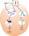
Non-canonical functions of intracellular complement. Intracellular complement activation in CD4+ T cells (and possibly other cells) occurs through the cleavage of intracellular C3 and C5 stores [or ‘imported’ hydrolyzed C3-C3(H2O)] by action of the C3-cleaving protease CTSL [for C3 and C3(H2O)] or a currently undefined protease for C5. The resulting C3a and C5a fragments engage their intracellular receptors (C3aR and C5aR1, respectively) and mediate mTOR activation and homeostatic survival (C3aR) and ROS production and possibly survival (C5aR1). Intracellular C3aR signaling occurs on lysosomes, although the intracellular compartment or compartments expressing C5aR1 are not yet defined. Abbreviations: mTOR, mechanistic target of rapamycin; ROS, reactive oxygen species; TCR, T cell receptor.
Figure 4
Figure 4
Direct effects of complement receptor activation on antigen presenting cells. On APCs (DCs), signaling of C5a and C3a through their receptors (C5aR1 and C3aR, respectively) upregulates MHC II and costimulatory B7 molecules and cAMP, ERK, and NF-κB signaling, leading to the secretion of IL-12, IL-23, IL-6, and/or TGF-β. TLR signals enhance secretion of C3a and C5a, which both act as ligands for their respective receptors and enhance receptor expression in a positive feedback loop. TLR signals, in conjunction with C5aR1 and C3aR signals, lead to the synergistic induction of IL-1β, IL-6, TNF-α, and IL-10, which are important cytokines for Th1 and Th17 induction. In human DCs and macrophages, CD46 stimulation also induces proinflammatory cytokine and NO production. C1q both positively and negatively regulates the inflammatory response, which depends partially upon the context/environment: C1q increases IL-1β and IL-12p70 and potentiates Th1 responses, whereas C1q bound to apoptotic cells inhibits IL-1β secretion and Th1 and Th17 proliferation. Additional anti-inflammatory signals are potentiated by CD55 (DAF), which reduces Th1 induction potential of APCs, and iC3b bound to CR3 results in TGF-β and IL-10 secretion and tolerance, which can be further regulated by Factor H and intracellular stores of iC3b. ‘Red’ arrows denote inhibitory activities of respective receptors. Abbreviations: APC, antigen-presenting cell; cAMP, cyclic adenosine monophosphate; DAF, decay-accelerating factor (CD55); DC, dendritic cell; ERK, extracellular signal–regulated kinase; MAC, membrane attack complex; NLRP3, NACHT-, leucine-rich repeat–, and pyrin domain–containing protein 3; NF-κB, nuclear factor kappa-light-chain enhancer of activated B cells; NO, nitric oxide; MHC II, major histocompatibility complex class II; MØ, macrophage; TLR, Toll-like receptor.
Figure 5
Figure 5
Direct effects of complement receptor signaling on human and mouse T cells. (a) In humans, activated T cells express C3aR and C5aR2 both intracellularly and extracellularly, whereas C5aR1 is expressed only observed intracellularly. Intracellular C5aR1 signaling results in mitochondrial ROS production and activation of the NLRP3 inflammasome, culminating in autocrine IL-1β signaling and enhanced Th1 function, which is inhibited by extracellular C5aR2 activation (through a yet-undefined mechanism). NLRP3 inflammasome activity and Th1 induction may be sustained by sublytic MAC formation on the T cell surface, with CD59 being a negative regulator of MAC formation and hence possibly of Th1 induction. C1q bound to opsonized immune complexes can be both a positive and a negative regulator of Th1 responses, as it regulates TNF-α and IFN-γ production in either a positive or a negative manner, depending upon the timing of the TCR signal. CR1 ligation inhibits IL-2 production and proliferation in activated T cells, whereas CR2 induces IL-8 production in RTEs. The potential signaling role of CD55 activation in human T cells is not understood. (b) In mice, activated T cells express surface C5aR1 (although its expression is controversial) and C3aR, but C5aR2 is expressed only intracellularly. C5aR1 and C3aR ligation results in PI3K and PKB activation, leading to increased IFN-γ and IL-2 production, increased IL-12Rβ expression and Th1 effector responses, and decreased FoxP3 Treg generation. Additionally, C5aR1 stimulation results in increased T cell survival through PKB activation. In contrast, C5aR2 and CD55 appear to be negative regulators of Th1 responses, where C5aR2 may induce TGF-β secretion and CD55 inhibits convertase formation. Mouse cells express a hybrid CR1/2 molecule, although its function is unknown. TCR and CD28 signals are not shown, as we focus on the signaling of complement receptors and regulators. ‘Red’ arrows denote inhibitory activities of receptors and ‘dashed’ lines denote observed effects with the underlying mechanisms not yet defined. Abbreviations: C5a-desArg, “desarginated” form of C5; MAC, membrane attack complex; NLRP3, NACHT-, leucine-rich repeat–, and pyrin domain–containing protein 3; PI3K, phosphatidylinositol-4,5-bisphosphate 3-kinase; PKB, protein kinase B; ROS, reactive oxygen species; RTEs, recent thymic immigrants; TLR, Toll-like receptor; Treg, regulatory T cell.
Figure 6
Figure 6
The role of the complosome in Th1 cell induction and contraction. (a) T cell receptor activation and CD28 costimulation of resting T cells induce the intracellular generation of the CD46 ligand C3b by CTSL cleavage and the increased expression of CD46 isoforms bearing CYT-1. Nuclear translocation of CYT-1 upon autocrine CD46 activation upregulates CD25 and CD132 gene expression, allowing for the enhanced IL-2 receptor signaling important for Th1 induction while decreasing CD127 gene expression. Autocrine CD46 CYT-1-driven signals also induce expression of the glucose transporter GLUT1 and the AA channel LAT1, allowing for increased glucose and AA influx. In parallel, CD46 CYT-1-mediated signals induce increased expression of LAMTOR5 and, via this the assembly of the lysosome-based mTORC1 machinery, which senses AA influx and drives the high levels of glycolysis and OXPHOS required for IFN-γ secretion. CD46-mediated signals also trigger increased intracellular C5a generation, which supports the mitochondrial metabolic activity and ROS production critical to normal Th1 induction. C5aR1-driven ROS production and mTORC1 activity also activate the NLRP3 inflammasome in TCR-stimulated T cells, a process that supports Th1 expansion via IL-1β functioning in an autocrine fashion. (b) During Th1 contraction and induction of IL-10 coexpression, CD46 isoform expression reverts to a CYT-2-predominant pattern (through an unknown mechanism), and this development, in conjunction with IL-2 receptor signaling (also through an unknown mechanism), results in reduced expression of GLUT1 and LAT1, downregulation of glycolysis and OXPHOS, and reduced IFN-γ production. Moreover, autocrine engagement of the surface-expressed C5aR2 via C5a or C5a-desArg secreted upon T cell activation contributes to negative regulation of mitochondrial activity and reduction of ROS (through inhibition of intracellular C5aR1 activity and/or a yet-undefined mechanism). ‘Red’ arrows denote inhibitory activities of receptors and ‘dashed’ lines denote observed effects with the underlying mechanisms not yet defined. Abbreviations: AA, amino acids; C5a-desArg, “desarginated” form of C5a; CTSL, cathepsin L; GLUT1, glucose transporter 1; LAMTOR5, late endosomal-lysosomal adaptor, MAPK, and mTOR activator 5; LAT1, neutral amino acid transporter 1; mTORC1, mechanistic target of rapamycin complex 1; NLRP3, NACHT-, leucine-rich repeat–, and pyrin domain–containing protein 3; OXPHOS, oxidative phosphorylation; ROS, reactive oxygen species; TCR, T cell receptor.
Figure 7
Figure 7
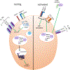
The role of the complosome in T cell homeostasis. In resting cells, intracellularly generated complement fragments sustain T cell self-regulation through the fine-tuning of basal metabolic activity via both. C3a generated by CTSL or the recycling pathway (shown in Figure 1c) and cell survival by C5a binding intracellular C5aR1 (through an unknown mechanism). This quiescent state is supported by complement-driven assembly of the IL-7R and by CD46 sequestration of Jagged-1, thus restraining Notch activity (left side of the cell). The brake imposed by CD46 and Jagged-1 is released in activated cells, when TCR stimulation drives CD46 shedding through metalloproteases, allowing for the release of Jagged-1 and, thus, increased Notch activity. The CD46-driven decrease in CD127 expression then also reduces incoming IL-7 signals (right side of the cell). ‘Dashed’ lines denote observed effects with the underlying mechanisms not yet defined. Abbreviations: CTSL, cathepsin L; mTORC1, mechanistic target of rapamycin complex 1; TCR, T cell receptor.
Figure 8
Figure 8
The evolution of complement from an intracellular driver of cell homeostasis and effector function to a serum sentinel system orchestrating host integrity. The ancient origin of complement and its coevolution and tight connection with basic cellular processes suggest that complement may have originally appeared as an intracellular system regulating single-cell homeostasis and may have functioned to mostly recognize and rectify danger derived from the self/within the cell (left). When life evolved into multicellular organisms, complement also became a secreted system and adopted an additional function of being a protector of the extracellular space against pathogens (right). Today, this dual role of complement likely explains why this ancient system in direct intra- and extracellular cross talk with other sensor and effector systems (such as TLRs, inflammasomes, and growth factors; not shown here) has such profound effects on normal cell and host function in immunity and beyond. Circles with solid outlines depict functions for complement that have been clearly demonstrated, whereas circles with dashed outlines are processes that we anticipate complement will contribute to. For many of the activities currently solely attributed to the liver-derived, extracellular complement, possible contributions from the intracellular complement system, the complosome, have not yet been assessed.

References

    1. Creagh EM, O’Neill LA. 2006. TLRs, NLRs and RLRs: a trinity of pathogen sensors that co-operate in innate immunity. Trends Immunol. 27:352–57 - PubMed
    1. Lavelle EC, Murphy C, O’Neill LA, Creagh EM. 2010. The role of TLRs, NLRs, and RLRs in mucosal innate immunity and homeostasis. Mucosal Immunol. 3:17–28 - PMC - PubMed
    1. Köhl J 2006. The role of complement in danger sensing and transmission. Immunol. Res. 34:157–76 - PubMed
    1. Walport M 2001. Complement. First of two parts. N. Engl. J. Med. 344:1058–66 - PubMed
    1. Walport M 2001. Complement. Second of two parts. N. Engl. J. Med. 344:1140–44 - PubMed

Publication types

Substances