Review
. 2018 Apr 26:36:461-488.
doi: 10.1146/annurev-immunol-042617-053019.

Unraveling the Complex Interplay Between T Cell Metabolism and Function

Affiliations
Review

Unraveling the Complex Interplay Between T Cell Metabolism and Function

Ramon I Klein Geltink et al. Annu Rev Immunol. .

Abstract

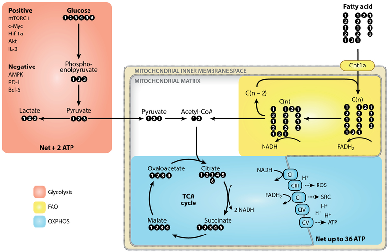
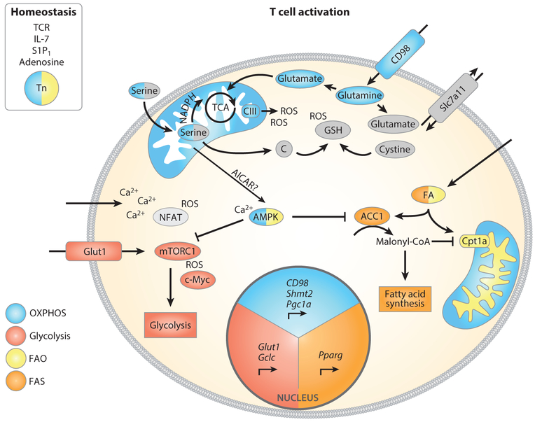
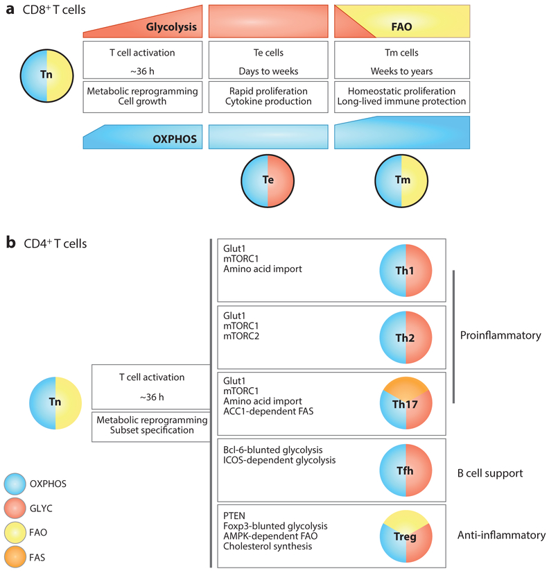
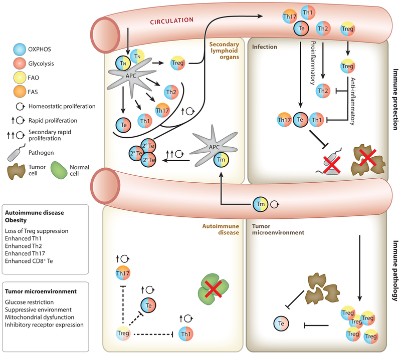
Metabolism drives function, on both an organismal and a cellular level. In T cell biology, metabolic remodeling is intrinsically linked to cellular development, activation, function, differentiation, and survival. After naive T cells are activated, increased demands for metabolic currency in the form of ATP, as well as biomass for cell growth, proliferation, and the production of effector molecules, are met by rewiring cellular metabolism. Consequently, pharmacological strategies are being developed to perturb or enhance selective metabolic processes that are skewed in immune-related pathologies. Here we review the most recent advances describing the metabolic changes that occur during the T cell lifecycle. We discuss how T cell metabolism can have profound effects on health and disease and where it might be a promising target to treat a variety of pathologies.

Keywords: T cell function; immunometabolism; immunotherapy; plasticity.


Figures

Figure 1
Figure 1
Core metabolic pathways for T cell survival, activation, differentiation, and function. Glucose is metabolized to pyruvate through the glycolysis pathway, a process controlled by many positive and negative factors. Pyruvate is converted to lactate and exported from the cell or transported into the mitochondria to feed into the TCA cycle. The TCA cycle reduces NAD+ to NADH, which is in turn oxidized by complexes of the electron transport chain. The transferred electrons are used to generate a proton gradient across the mitochondrial inner membrane. Complex V uses the proton-motive force of this gradient to generate ATP in a process called OXPHOS. Glycolysis nets 2 ATP, whereas OXPHOS can yield up to 36 ATP per molecule of glucose. Other substrates, such as fatty acids, can also feed into the TCA cycle. Cpt1a regulates the entrance of long-chain fatty acids into the mitochondria, where they are cleaved to generate acetyl-CoA. Abbreviations: AMPK, adenosine monophosphate–activated protein kinase; Bcl-6, B cell CLL/lymphoma 6; Hif-1α, hypoxia-inducible factor 1 alpha; mTORC1, mechanistic target of rapamycin complex 1; PD-1, programmed cell death protein 1; ROS, reactive oxygen species; TCA, tricarboxylic acid; OXPHOS, oxidative phosphorylation; SRC, spare respiratory capacity.
Figure 2
Figure 2
A variety of factors control Tn cell survival and metabolic pathway engagement after T cell activation. After TCR stimulation, mTORC1 and c-Myc promote glucose and amino acid uptake by enhancing expression of Glut1 and CD98. Shortly after T cell activation, mitochondrial biogenesis is engaged by Pgc1α. Cells also augment acquisition of serine, which is metabolized by mitochondrial Shmt2, and upregulated Gclc expression and cystine uptake support glutathione synthesis. Glutathione limits the accumulation of ROS from complex III, thereby facilitating T cell activation. Calcium import leads to activation of NFAT and AMPK, the latter of which might limit mTOR activation to preserve Tm cell differentiation potential. During the first division, asymmetric inheritance of the glycolytic modulators mTOR and c-Myc leads to differential T cell fate for the two daughter cells. Activated AMPK limits ACC1 activity, and increased expression of Pparg leads to upregulated fatty acid import, thereby modulating fatty acid metabolism after activation. Abbreviations: ACC1, acetyl-CoA carboxylase 1; AICAR, 5-aminoimidazole-4-carboxamide-1-β-D-ribofuranosyl 5’-monophosphate; AMPK, adenosine monophosphate–activated protein kinase; FA, fatty acid; FAO, fatty acid oxidation; FAS, fatty acid synthesis; Gclc, glutamate-cysteine ligase catalytic subunit; mTORC1, mechanistic target of rapamycin complex 1; NFAT, nuclear factor of activated T cells; OXPHOS, oxidative phosphorylation; Pgc1a, peroxisome proliferator–activated receptor gamma coactivator 1 alpha; Pparg, peroxisome proliferator–activated receptor gamma; ROS, reactive oxygen species; S1P1, sphingosine 1-phosphate; Shmt2, serine hypoxymethyltransferase 2; TCA, tricarboxylic acid (cycle); TCRT cell receptor; Tm, memory T cell; Tn, naive T cell.
Figure 3
Figure 3
Metabolic phenotypes of CD8+ and CD4+ T cell subsets. This schematic conveys the major pathways discussed in this review and is not exhaustive or exclusive of other pathways utilized by these T cell subtypes. (a) CD8+ Tn cells (outlined in black) rely on OXPHOS and use glucose to generate energy to maintain homeostatic proliferation and survival. Upon activation, CD8+ T cells increase OXPHOS and steadily increase their glycolytic rate, even before the first cell division. Fully differentiated CD8+ Te cells utilize glycolysis and OXPHOS. Tm cells decrease their glycolytic rate, coinciding with the engagement of FAO to fuel OXPHOS. Tm cells are endowed with more SRC than Tn cells, which underlies their rapid recall capacity upon challenge. (b) Like CD8+ Tn cells, CD4+ Tn cells (no outline) rely on OXPHOS and use glucose to generate energy to maintain homeostatic proliferation and survival. Upon activation, CD4+ T cells differentiate into diverse subsets with distinct roles and metabolic requirements. While all subsets engage glycolysis, Th2 and Th17 cells display high levels of glucose uptake. The master transcription factors of the Treg (Foxp3) and Tfh (Bcl-6) cell lineages repress glycolytic gene transcription, although depending on context Tregs also engage glycolysis. All subsets sustain OXPHOS, albeit at varying levels. Tregs engage FAO for suppressive function, whereas Th17 cells rely on de novo FAS. The balance of FAO and FAS contributes to the divergence between Th17 and Tregs. Abbreviations: ACC1, acetyl-CoA carboxylase 1; AMPK, adenosine monophosphate–activated protein kinase; Bcl-6, B-cell CLL/lymphoma 6; FAO, fatty acid oxidation; FAS, fatty acid synthesis; Foxp3, forkhead box P3; ICOS, inducible T cell costimulator; OXPHOS, oxidative phosphorylation; SRC, spare respiratory capacity; Te, effector T cell; Tfh, T follicular helper cell; Th, T helper cell; Tm, memory T cell; Tn, naive T cell; Treg, regulatory T cell.
Figure 4
Figure 4
T cell metabolism during an immune response. Tn cells are activated by mature APCs in secondary lymphoid organs (top left) and then proliferate and differentiate into Te cells that travel to peripheral tissues and mediate immune responses against pathogens or tumors. During an immune response Tregs regulate inflammation to limit tissue damage (top right). Upon resolution of an infection or tumor, long-lived CD8+ Tm cells (outlined in black) form and remain metabolically quiescent until they reencounter antigen and become reactivated, at which time they rapidly reengage glycolysis and promote OXPHOS (top left). Limited reliance on glycolysis allows Tregs to persist in the glucose-depleted tumor microenvironment, whereby they inhibit antitumor Te cell function. Te cell functions that require glycolytic engagement, such as the robust translation of IFN-γ, are also inhibited in the tumor microenvironment (bottom right). Cell-intrinsic disruptions to metabolic pathways in T cells, and systemic metabolic disease such as obesity, promote inappropriate T cell activation and differentiation (bottom left). Loss of suppressive function by Tregs leads to greater inflammatory function in CD4+ and CD8+ Te cells (outlined in black). In obesity, leptin signals during activation drive glycolysis and Th1/Th17 cells rather than Tregs. Abbreviations: APC, antigen-presenting cell; FAO, fatty acid oxidation; FAS, fatty acid synthesis; OXPHOS, oxidative phosphorylation; Te, effector T cell; Th, T helper cell; Tm, memory T cell; Tn, naive T cell; Treg, regulatory T cell.

References

    1. Weiner HL, Frenkel D. 2006. Immunology and immunotherapy of Alzheimer’s disease. Nat. Rev. Immunol 6:404–16 - PubMed
    1. Heppner FL, Ransohoff RM, Becher B. 2015. Immune attack: the role of inflammation in Alzheimer disease. Nat. Rev. Neurosci 16:358–72 - PubMed
    1. Saltiel AR, Olefsky JM. 2017. Inflammatory mechanisms linking obesity and metabolic disease. J. Clin. Investig 127:1–4 - PMC - PubMed
    1. Mathis D 2013. Immunological goings-on in visceral adipose tissue. Cell Metab. 17:851–59 - PMC - PubMed
    1. Warburg O 1925. The metabolism of carcinoma cells. J. Cancer Res 9:148–63

MeSH terms