. 2018 Apr 19;173(3):762-775.e16.
doi: 10.1016/j.cell.2018.03.076.

GPR68 Senses Flow and Is Essential for Vascular Physiology

Affiliations

GPR68 Senses Flow and Is Essential for Vascular Physiology

Jie Xu et al. Cell. .

Abstract

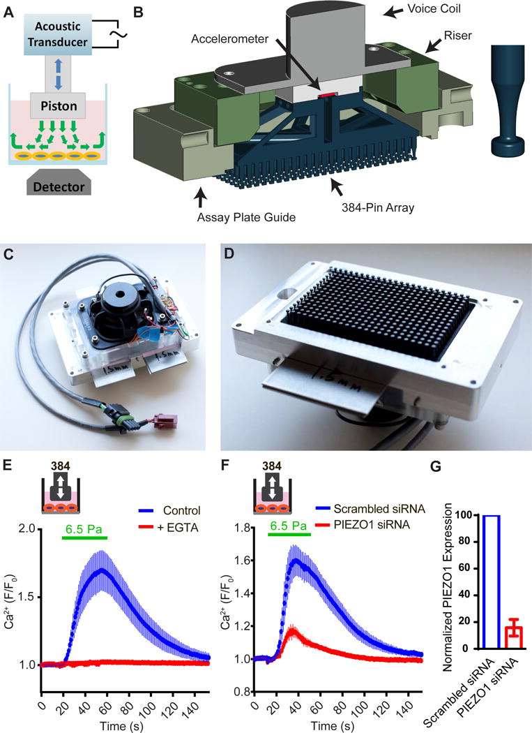
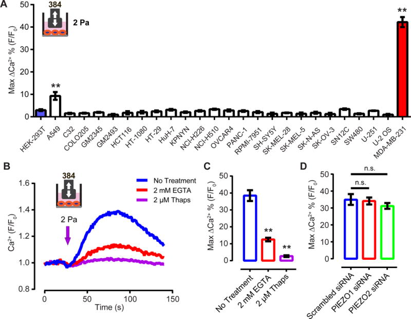
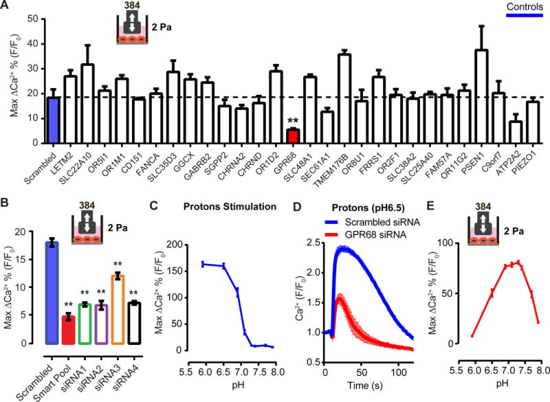
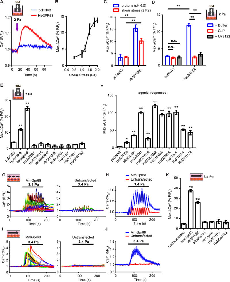
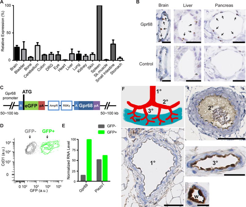
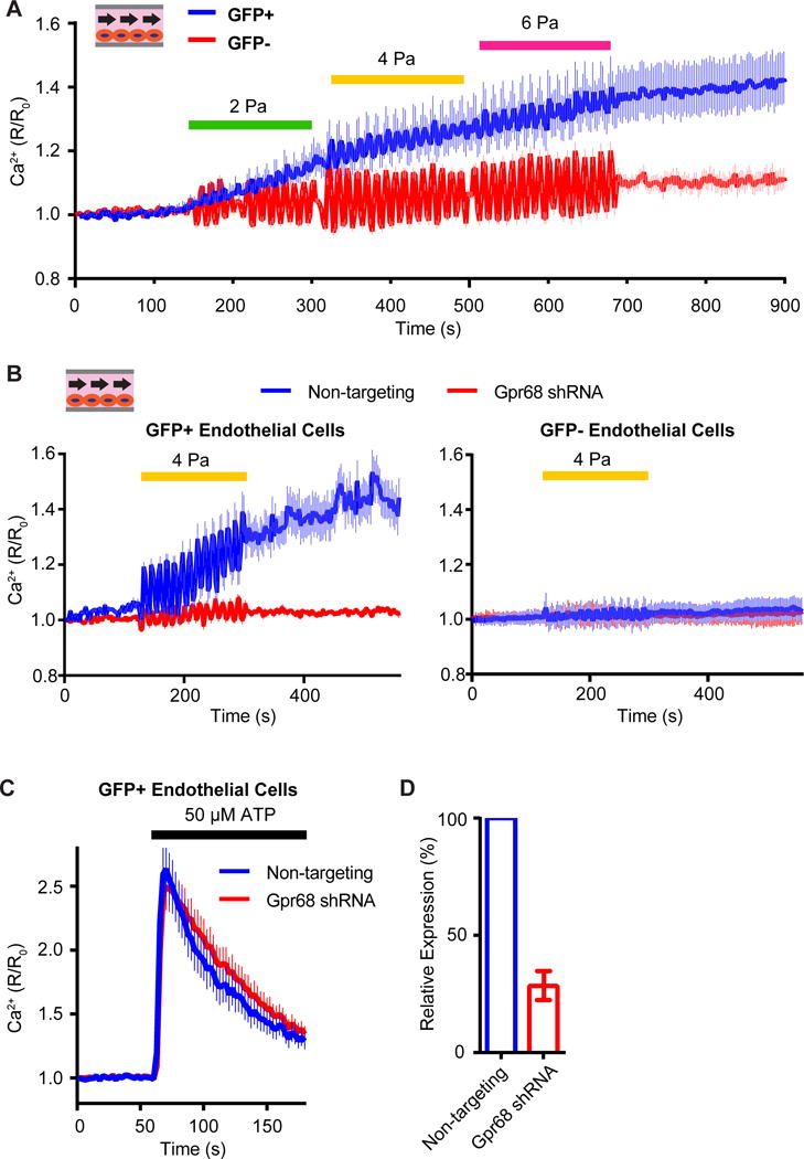
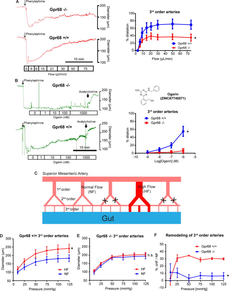
Mechanotransduction plays a crucial role in vascular biology. One example of this is the local regulation of vascular resistance via flow-mediated dilation (FMD). Impairment of this process is a hallmark of endothelial dysfunction and a precursor to a wide array of vascular diseases, such as hypertension and atherosclerosis. Yet the molecules responsible for sensing flow (shear stress) within endothelial cells remain largely unknown. We designed a 384-well screening system that applies shear stress on cultured cells. We identified a mechanosensitive cell line that exhibits shear stress-activated calcium transients, screened a focused RNAi library, and identified GPR68 as necessary and sufficient for shear stress responses. GPR68 is expressed in endothelial cells of small-diameter (resistance) arteries. Importantly, Gpr68-deficient mice display markedly impaired acute FMD and chronic flow-mediated outward remodeling in mesenteric arterioles. Therefore, GPR68 is an essential flow sensor in arteriolar endothelium and is a critical signaling component in cardiovascular pathophysiology.

Keywords: GPCR; blood flow; mechanosensation; mechanotransduction; outward remodeling; shear stress; vascular biology; vasodilation.


Conflict of interest statement

DECLARATION OF INTERESTS

The authors declare no competing interests.

Figures

Figure 1
Figure 1. Construction and validation of 384-well high-throughput shear stress stimulation system
(A) Schematic representation of the disturbed flow stimulation system. (B) Left, drawing depicts the major components. Right, the bubble-relief geometry of the stimulator pins. (C) Top view showing the acoustic transducer assembly. (D) Bottom view showing the 384-pin array. (E) Intracellular calcium levels of HeLa cells measured by FLIPR, under 6.5 Pa shear stress stimulation in the presence of 2.5 mM EGTA or untreated control. The viscosity of the assay buffer was increased to 8.37×10⁻³ Pa·s by adding PVP to 2% (w/w). Disturbed flow (0.2 s on, 2 s off) was applied at 60 Hz for 40 s. Data was average of 48 wells from 3 trials for each condition. (E) Intracellular calcium levels of HeLa cells under the same disturbed flow stimulation after the treatment of scrambled siRNA or hPIEZO1 siRNA. Data is average of 48 wells from 3 trials for each condition. (F) Relative mRNA level of PIEZO1 in HUVECs transfected with PIEZO1 siRNA compared with cells treated with scrambled siRNA (n=3).
Figure 2
Figure 2. MDA-MB-231 cells show PIEZO1- and PIEZO2-independent shear stress-induced calcium transients
(A) Responses of various human cancer cell lines to shear stress at 2 Pa, 60 Hz, with 0.2 s on, 2s off for 40 s. ** p<0.01 (B) The response of MDA-MB-231 cells to shear stress at 2 Pa, measured by FLIPR. The shear stress was applied for 4 s at 60 Hz. EGTA was added to the cells 2 min prior to the onset of shear stress. Thapsigargin was incubated with cells for 15 min before the shear stress stimulation. (C) Quantification of the MDA-MB-231 cells’ response to shear stress in the presence of the EGTA and thapsigargin. ** p<0.01. (D) Quantification of the MDA-MB-231 cells’ response to shear stress 72 h after transfection of PIEZO1 and PIEZO2 siRNA. n.s., not significant.
Figure 3
Figure 3. GPR68 is necessary for shear stress-induced calcium transients in MDA-MB-231 cells
(A)The response of MDA-MB-231 cells after treatment of siRNAs against the indicated candidates. ** p<0.01 (B) Quantification of the MDA-MB-231 cells’ response to shear stress after treatment with smartpool siRNA oligos and the individual siRNA oligos against GPR68. ** p<0.01. (C) Response of MDA-MB-231 cells to proton stimulation at various final pH. The cells were incubated with HBSS at pH7.4 before the addition of the HBSS with various acidity. n=3. (D) The response of MDA-MB-231 cells to proton stimulation (final pH 6.5) after knocking down GPR68 by siRNA smartpool. (E) The response of MDA-MB-231 cells to shear stress stimulation in buffers with various pH. The cells were incubated with assay buffer at pH7.4. The pH was then changed by adding buffer with various acidity 3 min prior to the onset of shear stress stimulation.
Figure 4
Figure 4. GPR68 is sensitive to shear stress imposed by both disturbed and laminar flow in HEK-293T cells
(A) Disturbed flow-induced calcium transients in HEK-293T cells transfected with human GPR68 or vector control 48 h before the assay. Flow was applied at 60 Hz for 4s (arrow indicates the onset of the flow). (B) Quantification of the amplitude of the calcium transients evoked by disturbed shear stress of increasing intensities. n=3 wells per intensity tested. (C) Response of HEK-293T cells to pH 6.5 stimulation and 2 Pa disturbed shear stress. ** p<0.01. (D) Response of HEK-293T cells to 2 Pa disturbed flow in the presence of 20 μM Cu2+ or 10 μM U73122. Cu2+ or U73122 was added to the assay buffer 2 min prior to the start of the flow. n.s., not significant. ** p<0.01. (E) The shear stress induced responses of HEK-293T cells transiently-transfected with various human and mouse GPCRs. ** p<0.01. (F) The agonist response of HEK-293T cells transiently-transfected with various human and mouse GPCRs. ** p<0.01. The chemical activators used were: pH 6.5 for human and murine GPR68, 0.5 μM Angiotensin II for AGTR1, 0.5 μM [Arg8]-Vasopressin for AVPR1A, 0.5 μM Bradykinin for BDKRB2, 0.5 μM Acetylcholine Chloride for CHRM5, 0.5 μM Endothelin I for EDNRA, 0.5 μM Histamine Dihydrochloride for HRH1, 20 μM Parathyroid Hormone (1-34) for PTHR1, and 100 mM lactate for GPR132. (G) Representative traces of intracellular calcium levels in HEK-293T cells transfected with mouse Gpr68-IRES-eGFP upon pulsatile laminar flow stimulation. Cells with GFP signal were considered transfected and the GFP negative ones were untransfected. Pulsatile laminar flow was applied at 3.4 Pa, 1 Hz for 120 s. (H) Quantification of the response of HEK cells to pulsatile laminar flow. n=172 for MmGpr68 transfected and 165 for untransfected. (I) Representative traces of intracellular calcium levels in HEK-293T cells transfected with mouse Gpr68-IRES-eGFP upon steady laminar flow stimulation. Steady laminar flow was applied at 3.4 Pa for 120 s. (J) Response of HEK cells to steady laminar flow. n=162 for MmGpr68 transfected and 183 for untransfected. (K) Responses of HEK-293T cells transfected with various putative mechanosensors to steady laminar flow at 3.4 Pa. ** p<0.01.
Figure 5
Figure 5. GPR68 expression is detected in endothelial cells of small-diameter vessels
(A) Murine Gpr68 mRNA expression profile determined by qRT-PCR. Gapdh was used as the reference gene. The expression levels were normalized to spleen. n=2~3. (B) Representative images of colorimetric RNAscope in situ hybridization for Gpr68 vascular endothelial cells of small diameter blood vessels in brain, pancreas and liver. Scale bars: 25 μm. Arrows indicate cells with positive signal. (C) Schematic diagram of the BAC transgenic construct that is integrated in the genome of Gpr68 eGFP reporter mice. The blue A-box is the homologous sequence that guides recombination and insert eGFP after the start codon of Gpr68. Poly-A sequence following the eGFP blocks the transcription of the downstream genes within the construct (including Gpr68). pA: poly-A sequence; AmpR, amplicilin-resistance genes; R6Kγ, origin of DNA replication (AmpR and R6Kγ are used in the initial selection of BAC constructs). (D) FACS plot of primary endothelial cells isolated from the bladder of Gpr68-eGFP reporter mice. Cells that are CD31+ and CD45− are shown, and grouped in the GFP+ and GFP− populations. (E) Normalized RNA levels (RPKM) of Gpr68 and Piezo1 from the RNAseq data of GFP+ and GFP− endothelial cells. Bladder cells from 3 batches of isolation and sorting (10 mice in total) were pooled and subjected to RNAseq. (F) Representative images of GFP antibody staining in arteries of 1st order (1°), 2nd order (2°), 3rd order (3°) superior mesenteric vessels, and the vessels in the wall of small intestine (w). Scale bars: 50 μm for 1° and 2° vessels, 25 μm for 3° vessels, and 10 μm for vessels in the small intestine wall. 8 groups of vessels from 4 mice were examined.
Figure 6
Figure 6. Gpr68 is necessary for laminar flow-induced calcium transients in murine primary microvascular endothelial cells
(A) Responses of mouse primary microvascular endothelial cells (MVECs) to shear stress imposed by pulsatile laminar flow with increasing amplitudes. The MVECs were isolated from cerebrum of the Gpr68 eGFP reporter mice. Pulsatile laminar flow of different amplitudes are applied at 1 Hz for 180s. n=148 (GFP+) and 155 (GFP−). Data is pooled from 3 trials. (B) Responses of GFP+ and GFP− MVECs to pulsatile laminar shear stress at 4 Pa. The MVECs were infected with lentivirus containing non-targeting shRNA or Gpr68 shRNA constructs. The pulsatile flow is applied at 1 Hz for 180 s. n=166 (non-targeting) and 173 (Gpr68 shRNA). 3 trials. (C) The calcium transients in primary MVECs induced by to 50 μM ATP. n=223 (non-targeting) and 198 (Gpr68 shRNA). 3 trials. (D) Quantification of the knockdown efficiency of lentiviral shRNA against Gpr68. n=3.
Figure 7
Figure 7. Gpr68 is required for flow-mediated dilation and outward remodeling in third order mesenteric arteries (MAs)
(A) Flow-mediated dilation (FMD) response of third order MAs isolated from Gpr68 −/− mice and WT littermates. Vessels were cannulated and pre-constricted using 1 μM phenylephrine, and subjected to stepwise increases in flow rates. Left, representative recordings of vessel diameter change. Scale bar: 10 minutes. Right, quantification of FMD response in 3rd order MAs. p=0.044, two way ANOVA, n=9 for Gpr68 KO and n=7 for WT. (B) Dilation responses of third order MAs to Ogerin. Vessels were cannulated and pre-constricted using 1 μM phenylephrine, and subjected to Ogerin with increasing concentration. Left, representative recordings of vessel diameter change. Scale bar: 10 minutes. Right, quantification of Ogerin-induced dilation 3rd order MAs. Vessels from 6 KO and 6 WT mice were tested. P=0.018, two way ANOVA. Inset, structure of Ogerin. (C) Schematic representation of the surgery applied to mesenteric arteries to create local high flow (HF) and normal (NF) regions (see EXPERIMENTAL PROCEDURE). (D, E) Flow-mediated remodeling (FMR) of 3rd order mesenteric arteries isolated from WT (P=0.0045) and Gpr68 KO mice (P=0.95). (F) The extent of outward remodeling as indicated by the percentage of vessel diameter increase (*P=0.0309). *p<0.05, two way ANOVA for repeated measures, n=5 for Gpr68 −/− and n=7 for WT.

Comment in

Similar articles

Cited by

References

    1. Aoki H, Mogi C, Hisada T, Nakakura T, Kamide Y, Ichimonji I, Tomura H, Tobo M, Sato K, Tsurumaki H, et al. Proton-sensing ovarian cancer G protein-coupled receptor 1 on dendritic cells is required for airway responses in a murine asthma model. PloS one. 2013;8:e79985. - PMC - PubMed
    1. Burns ME, Pugh EN., Jr Lessons from photoreceptors: turning off g-protein signaling in living cells. Physiology. 2010;25:72–84. - PMC - PubMed
    1. Caillon A, Grenier C, Grimaud L, Vessieres E, Guihot AL, Blanchard S, Lelievre E, Chabbert M, Foucher ED, Jeannin P, et al. The angiotensin II type 2 receptor activates flow-mediated outward remodelling through T cells-dependent interleukin-17 production. Cardiovascular research. 2016;112:515–525. - PubMed
    1. Chachisvilis M, Zhang YL, Frangos JA. G protein-coupled receptors sense fluid shear stress in endothelial cells. Proceedings of the National Academy of Sciences of the United States of America. 2006;103:15463–15468. - PMC - PubMed
    1. Chen P, Zuo H, Xiong H, Kolar MJ, Chu Q, Saghatelian A, Siegwart DJ, Wan Y. Gpr132 sensing of lactate mediates tumor-macrophage interplay to promote breast cancer metastasis. Proceedings of the National Academy of Sciences of the United States of America. 2017;114:580–585. - PMC - PubMed

Publication types

MeSH terms