. 2018 Apr 20;17(1):90.
doi: 10.1186/s12944-018-0740-6.

A20 deubiquitinase controls PGC-1α expression in the adipose tissue

Affiliations

A20 deubiquitinase controls PGC-1α expression in the adipose tissue

Bruna Bombassaro et al. Lipids Health Dis. .

Abstract

Background: Peroxisome proliferator-activated receptor γ coactivator- 1alpha (PGC-1α) plays an important role in whole body metabolism and, particularly in glucose homeostasis. Its expression is highly regulated and, small variations in tissue levels can have a major impact in a number of physiological and pathological conditions. Recent studies have shown that the ubiquitin/proteasome system plays a role in the control of PGC-1α degradation.

Methods: Here we evaluated the interaction of PGC-1α with the protein A20, which plays a dual-role in the control of the ubiquitin/proteasome system acting as a deubiquitinase and as an E3 ligase. We employed immunoprecipitation, quantitative real-time PCR and immunofluorescence staining to evaluate PGC-1α, A20, PPARγ and ubiquitin in the adipose tissue of humans and mice.

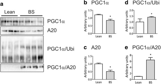
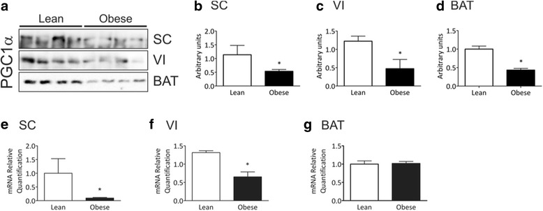
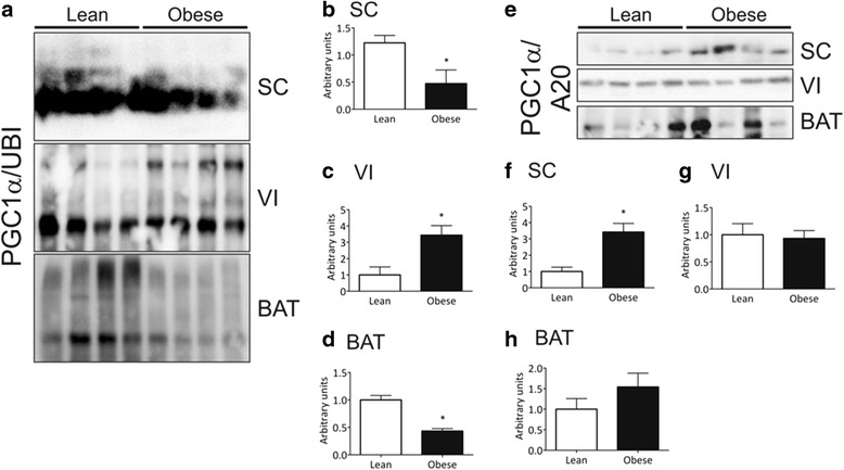
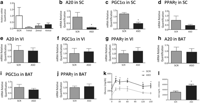
Results: In distinct sites of the adipose tissue, A20 binds to PGC-1α. At least in the subcutaneous fat of humans and mice the levels of PGC-1α decrease during obesity, while its physical association with A20 increases. The inhibition of A20 leads to a reduction of PGC-1α and PPARγ expression, suggesting that A20 acts as a protective factor against PGC-1α disposal.

Conclusion: We provide evidence that mechanisms regulating PGC-1α ubiquitination are potentially involved in the control of the function of this transcriptional co-activator.

Keywords: Diet; Fat; Glucose; Obesity; Ubiquitination.


Conflict of interest statement

Ethics approval and consent to participate

The human part of the study was approved by the University of Campinas Ethics Committee for Medical Research (#833/2010). The experimental part of the study were performed in accordance with the guidelines of the Brazilian College for Animal Experimentation and were approved by the University of Campinas Ethics Committee (#CEUA 2216–1).

Consent for publication

All authors agree with this publication.

Competing interests

The authors declare that they have no competing interests.

Publisher’s Note

Springer Nature remains neutral with regard to jurisdictional claims in published maps and institutional affiliations.

Figures

Fig. 1
Fig. 1
PGC-1α and A20 expression in the adipose tissue of humans. Samples containing 500 μg total protein from abdominal subcutaneous adipose tissue specimens, collected from 9 lean volunteers and 12 obese subjects during a Roux-in-Y gastric bypass (BS) were used in immunoprecipitation experiments employing anti-PGC-1α (a, b, d and e) or anti-A20 (a and c) as primary antibodies. The immunoprecipitation procedure used antibodies sufficient to immunodeplete the sample. The immunoprecipitates were separated by SDS-PAGE, transferred to nitrocellulose membranes and blotted with anti-PGC-1α (a and b), anti-A20 (a, c and e) or anti-ubiquitin (a and d) antibodies. In A, typical blots are depicted; in B-E the quantification of bands is graphically represented. *p < 0.05 vs. lean
Fig. 2
Fig. 2
PGC-1α expression in distinct adipose tissue depots of mice. Samples containing 500 μg total protein (a-d) or 25 ng cDNA (E-G) from subcutaneous (SC), visceral (VI) or brown (BAT) adipose tissue depots from lean or obese mice were used in immunoprecipitation (a-d) or quantitative real-time PCR (e-g) experiments. In a-d, samples were immunoprecipitated employing anti-PGC-1α as primary antibody. The immunoprecipitation procedure used antibodies sufficient to immunodeplete the sample. The immunoprecipitates were separated by SDS-PAGE, transferred to nitrocellulose membranes and blotted with anti-PGC-1α antibody. In a, typical blots are depicted; in b-d the quantification of bands is graphically represented. In e-g, the quantification of PGC-1α mRNA obtained by quantitative real-time PCR is represented graphically. In all experiments n = 5–6, *p < 0.05 vs. lean
Fig. 3
Fig. 3
A20 expression in distinct adipose tissue depots of mice. Samples containing 500 μg total protein (a-d) or 25 ng cDNA (e-g) from subcutaneous (SC), visceral (VI) or brown (BAT) adipose tissue depots from lean or obese mice were used in immunoprecipitation (a-d) or quantitative real-time PCR (e-g) experiments. In a-d, samples were immunoprecipitated employing anti-A20 as primary antibody. The immunoprecipitation procedure used antibodies sufficient to immunodeplete the sample. The immunoprecipitates were separated by SDS-PAGE, transferred to nitrocellulose membranes and blotted with anti-A20 antibody. In a, typical blots are depicted; in b-d the quantification of bands is graphically represented. In e-g, the quantification of A20 mRNA obtained by quantitative real-time PCR is represented graphically. In all experiments n = 6, *p < 0.05 vs. lean
Fig. 4
Fig. 4
PGC-1α association with ubiquitin and A20 in distinct adipose tissue depots of mice. Samples containing 500 μg total protein from subcutaneous (SC), visceral (VI) or brown (BAT) adipose tissue depots from lean or obese mice were used in immunoprecipitation experiments employing anti-PGC-1α as primary antibody. The immunoprecipitation procedure used antibodies sufficient to immunodeplete the sample. The immunoprecipitates were separated by SDS-PAGE, transferred to nitrocellulose membranes and blotted with anti-ubiquitin (a-d) or anti-A20 (e-h) antibodies. In a and e, typical blots are depicted; in b-d and f-h the quantification of bands is graphically represented. In all experiments n = 5–6, *p < 0.05 vs. lean
Fig. 5
Fig. 5
Immunofluoescence evaluation of the co-localizations of PGC-1α with ubiquitin and A20. Subcutaneous (SC), visceral (VI) or brown (BAT) adipose tissue specimens from obese mice were used in immunofluorescence assays to detect the presence and co-localizations of PGC-1α and ubiquitin (Ubi) (a) and PGC-1α and A20 (b). Figures are typical representations obtained from a total of two distinct experiments
Fig. 6
Fig. 6
Inhibiting A20 expression. Obese Swiss mice were treated once a day, for 7 days, with a single intraperitoneal injection of a 100 μl solution containing either A20 antisense (ASO) or scrambled (SCR) oligonucleotide. Fragments from subcutaneous (SC) (a-d), visceral (VI) (e-g) or brown (BAT) (h-j) adipose tissue were employed in quantitative real-time PCR experiments. In a, A20 transcript expression was evaluated in a dose-response experiment to determine the efficiency of the method. In b-l, the doses of ASO and SCR employed were always 2 nmol in 100 μl/dose. In b-j, the expressions of A20 (b, e, h); PGC-1α (c, f, i); and PPARγ (d, g, j) were determined and are represented graphically. The blood glucose levels during a glucose tolerance test (k) and the respective area under the glucose curve (l) are represented graphically. In all experiments n = 8; *p < 0.05 vs. SCR

References

    1. Handschin C, Spiegelman BM. Peroxisome proliferator-activated receptor gamma coactivator 1 coactivators, energy homeostasis, and metabolism. Endocr Rev. 2006;27:728–735. doi: 10.1210/er.2006-0037. - DOI - PubMed
    1. Fuentes E, Guzman-Jofre L, Moore-Carrasco R, Palomo I. Role of PPARs in inflammatory processes associated with metabolic syndrome (review) Mol Med Rep. 2013;8:1611–1616. doi: 10.3892/mmr.2013.1714. - DOI - PubMed
    1. Puigserver P, Wu Z, Park CW, Graves R, Wright M, Spiegelman BM. A cold-inducible coactivator of nuclear receptors linked to adaptive thermogenesis. Cell. 1998;92:829–839. doi: 10.1016/S0092-8674(00)81410-5. - DOI - PubMed
    1. Bargut TCL, Souza-Mello V, Aguila MB, Mandarim-de-Lacerda CA: Browning of white adipose tissue: lessons from experimental models. Horm Mol Biol Clin Investig 2017;31:1–13. - PubMed
    1. De Souza CT, Gasparetti AL, Pereira-da-Silva M, Araujo EP, Carvalheira JB, Saad MJ, Boschero AC, Carneiro EM, Velloso LA. Peroxisome proliferator-activated receptor gamma coactivator-1-dependent uncoupling protein-2 expression in pancreatic islets of rats: a novel pathway for neural control of insulin secretion. Diabetologia. 2003;46:1522–1531. doi: 10.1007/s00125-003-1222-5. - DOI - PubMed

MeSH terms

Substances