. 2018 May 1;121(9):1081-1089.
doi: 10.1016/j.amjcard.2018.01.022. Epub 2018 Feb 6.

Stress Myocardial Blood Flow Heterogeneity Is a Positron Emission Tomography Biomarker of Ventricular Arrhythmias in Patients With Hypertrophic Cardiomyopathy

Affiliations

Stress Myocardial Blood Flow Heterogeneity Is a Positron Emission Tomography Biomarker of Ventricular Arrhythmias in Patients With Hypertrophic Cardiomyopathy

Dai-Yin Lu et al. Am J Cardiol. .

Abstract

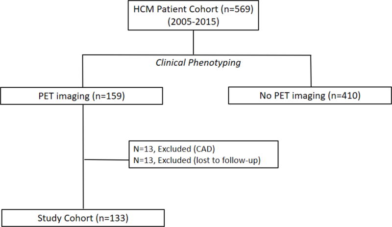
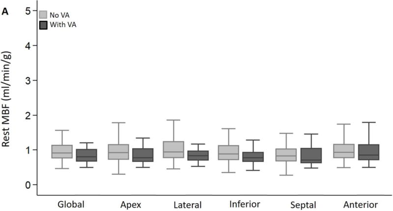
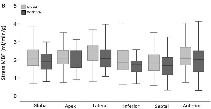
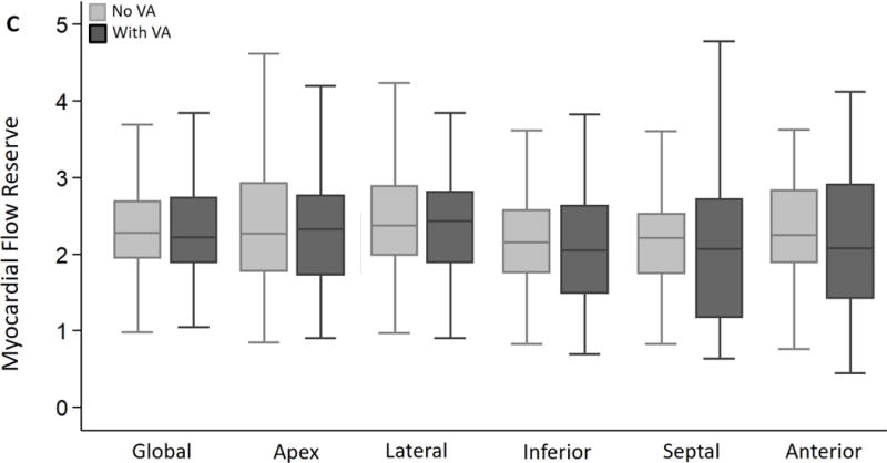
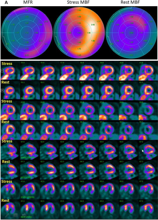
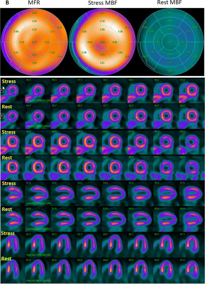
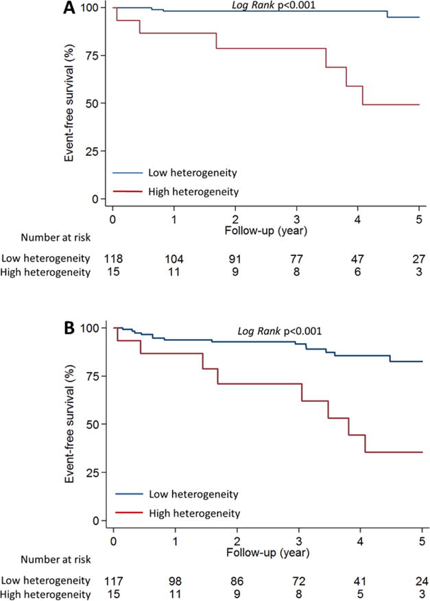
Patients with hypertrophic cardiomyopathy (HC) are at increased risk of sudden cardiac death. Abnormalities in myocardial blood flow (MBF) detected by positron emission tomography (PET) are common in HC, but a PET marker that identifies patients at risk of sudden cardiac death is lacking. We hypothesized that disparities in regional myocardial perfusion detected by PET would identify patients with HC at risk of ventricular arrhythmias. To test this hypothesis, we quantified global and regional MBFs by 13NH3-PET at rest and at stress, and developed a heterogeneity index to assess MBF heterogeneity in 133 symptomatic patients with HC. The MBF heterogeneity index was computed by dividing the highest by the lowest regional MBF value, at rest and after vasodilator stress, in each patient. High stress MBF heterogeneity was defined as an index of ≧1.85. Patients with HC were stratified by the presence or the absence of ventricular arrhythmias, defined as sustained ventricular tachycardia (VT) and/or nonsustained VT, during follow-up. We found that global and regional MBFs at rest and stress were similar in patients with HC with or without ventricular arrhythmias. Variability in regional stress MBF was observed in both groups, but the stress MBF heterogeneity index was significantly higher in patients with HC who developed ventricular arrhythmias (1.82 ± 0.77 vs 1.49 ± 0.25, p <0.001). A stress MBF heterogeneity index of ≧1.85 was an independent predictor of both sustained VT (hazard ratio 16.1, 95% confidence interval 3.2 to 80.3) and all-VT (sustained-VT + nonsustained VT: hazard ratio 3.7, 95% confidence interval 1.4 to 9.7). High heterogeneity of stress MBF, reflected by an MBF heterogeneity index of ≥1.85, is a PET biomarker for ventricular arrhythmias in symptomatic patients with HC.


Conflict of interest statement

Conflict of interest: None

Figures

Figure 1
Figure 1. Flow chart for patient inclusion in HC-PET study
Figure 2
Figure 2. Global and regional MBF and MFR in HC patients stratified by presence or absence of ventricular arrhythmias (VA) during follow up
(A) Global and regional MBF at rest, (B) Global and regional MBF during vasodilator stress and (C) Global and regional MFR, were similar between HC patients who developed ventricular arrhythmias (sustained VT, NSVT) and HC patients who had no evidence of ventricular arrhythmias during follow up.
Figure 3
Figure 3. Regional distribution of stress MBF in HC cohort
The lateral wall demonstrated the highest stress MBF in 56% of patients, and was followed by the anterior wall and apex, respectively, whereas the septum exhibited the lowest stress MBF in ~44% of patients.
Figure 4
Figure 4. Rest and stress MBF heterogeneity index stratified by presence or absence of ventricular arrhythmias (VA)
The rest MBF heterogeneity index was similar in patients with/without ventricular arrhythmias. HC patients who developed sustained ventricular arrhythmias (sustained VT, NSVT) had a significantly higher stress MBF heterogeneity index than patients who did not have ventricular arrhythmias during follow up.
Figure 5
Figure 5. 13NH3-PET polar plots and perfusion images
(A) 13NH3-PET polar plots and perfusion images in HC patient with stress MBF heterogeneity index of 3.12, who developed sustained VT during follow up. Polar plots demonstrate evidence of mild reduction of MFR globally, and severe reduction of MFR in the septum. Perfusion images reveal reversible myocardial ischemia in anterior and septal regions (SDS=7). (B) 13NH3-PET polar plots and perfusion images in HC patient with stress MBF heterogeneity index of 1.07, who did not develop ventricular arrhythmias during follow up. MFR is normal and there is no evidence of vasodilator-induced myocardial ischemia (SDS=1). MBF: myocardial blood flow, MFR: myocardial flow reserve.
Figure 6
Figure 6. Kaplan-Meier curves comparing ventricular arrhythmia outcome stratified by the stress MBF heterogeneity index
HC patients with stress MBF heterogeneity index ≥1.85 (red line) had significantly higher risk of developing ventricular arrhythmias, characterized by (A) sustained-VT, as well as (B) All-VT (sustained-VT+NSVT), when compared to HC patients with stress MBF heterogeneity index <1.85 (blue line).

References

    1. Gersh BJ, Maron BJ, Bonow RO, Dearani JA, Fifer MA, Link MS, Naidu SS, Nishimura RA, Ommen SR, Rakowski H, Seidman CE, Towbin JA, Udelson JE, Yancy CW, American College of Cardiology Foundation/American Heart Association Task Force on Practice G 2011 ACCF/AHA Guideline for the Diagnosis and Treatment of Hypertrophic Cardiomyopathy: a report of the American College of Cardiology Foundation/American Heart Association Task Force on Practice Guidelines. Developed in collaboration with the American Association for Thoracic Surgery, American Society of Echocardiography, American Society of Nuclear Cardiology, Heart Failure Society of America, Heart Rhythm Society, Society for Cardiovascular Angiography and Interventions, and Society of Thoracic Surgeons. J Am Coll Cardiol. 2011;58:e212–260. - PubMed
    1. Jung WI, Sieverding L, Breuer J, Hoess T, Widmaier S, Schmidt O, Bunse M, van Erckelens F, Apitz J, Lutz O, Dietze GJ. 31P NMR spectroscopy detects metabolic abnormalities in asymptomatic patients with hypertrophic cardiomyopathy. Circulation. 1998;97:2536–2542. - PubMed
    1. Abraham MR, Bottomley PA, Dimaano VL, Pinheiro A, Steinberg A, Traill TA, Abraham TP, Weiss RG. Creatine kinase adenosine triphosphate and phosphocreatine energy supply in a single kindred of patients with hypertrophic cardiomyopathy. Am J Cardiol. 2013;112:861–866. - PMC - PubMed
    1. Dass S, Cochlin LE, Suttie JJ, Holloway CJ, Rider OJ, Carden L, Tyler DJ, Karamitsos TD, Clarke K, Neubauer S, Watkins H. Exacerbation of cardiac energetic impairment during exercise in hypertrophic cardiomyopathy: a potential mechanism for diastolic dysfunction. Eur Heart J. 2015;36:1547–1554. - PubMed
    1. Cecchi F, Olivotto I, Gistri R, Lorenzoni R, Chiriatti G, Camici PG. Coronary microvascular dysfunction and prognosis in hypertrophic cardiomyopathy. N Engl J Med. 2003;349:1027–1035. - PubMed

Publication types

MeSH terms