. 2018 May:11:113-128.
doi: 10.1016/j.molmet.2018.03.006. Epub 2018 Mar 15.

Growth hormone receptor-deficient pigs resemble the pathophysiology of human Laron syndrome and reveal altered activation of signaling cascades in the liver

Affiliations

Growth hormone receptor-deficient pigs resemble the pathophysiology of human Laron syndrome and reveal altered activation of signaling cascades in the liver

Arne Hinrichs et al. Mol Metab. 2018 May.

Abstract

Objective: Laron syndrome (LS) is a rare, autosomal recessive disorder in humans caused by loss-of-function mutations of the growth hormone receptor (GHR) gene. To establish a large animal model for LS, pigs with GHR knockout (KO) mutations were generated and characterized.

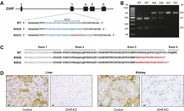
Methods: CRISPR/Cas9 technology was applied to mutate exon 3 of the GHR gene in porcine zygotes. Two heterozygous founder sows with a 1-bp or 7-bp insertion in GHR exon 3 were obtained, and their heterozygous F1 offspring were intercrossed to produce GHR-KO, heterozygous GHR mutant, and wild-type pigs. Since the latter two groups were not significantly different in any parameter investigated, they were pooled as the GHR expressing control group. The characterization program included body and organ growth, body composition, endocrine and clinical-chemical parameters, as well as signaling studies in liver tissue.

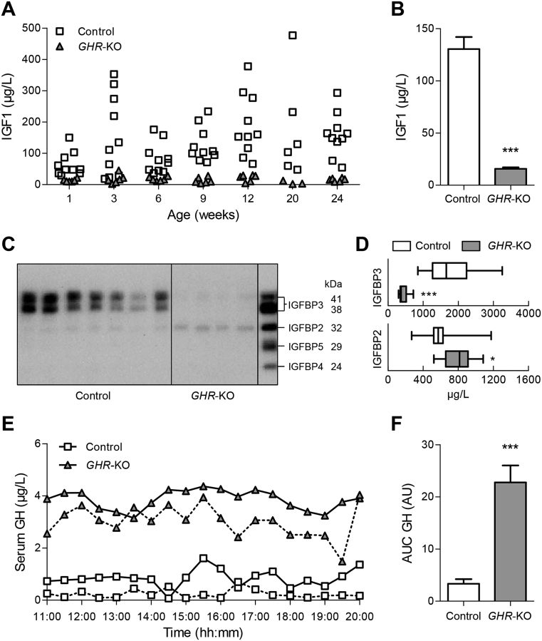
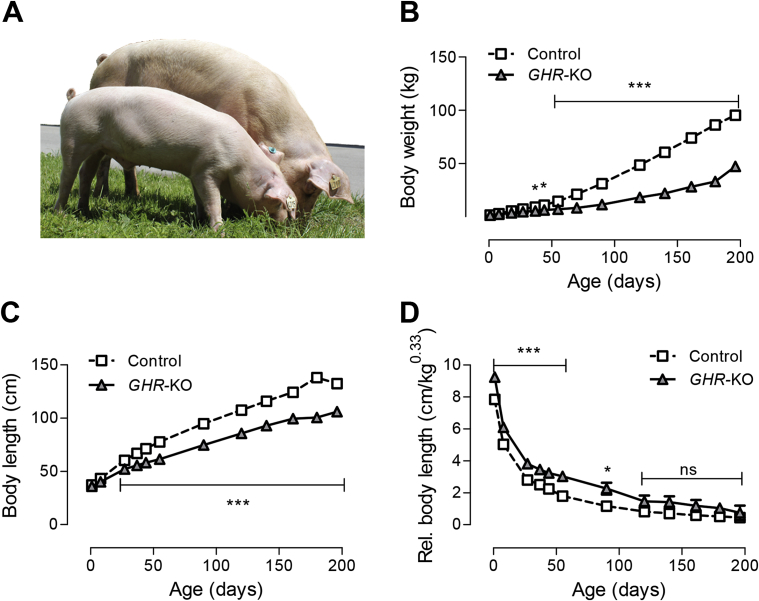
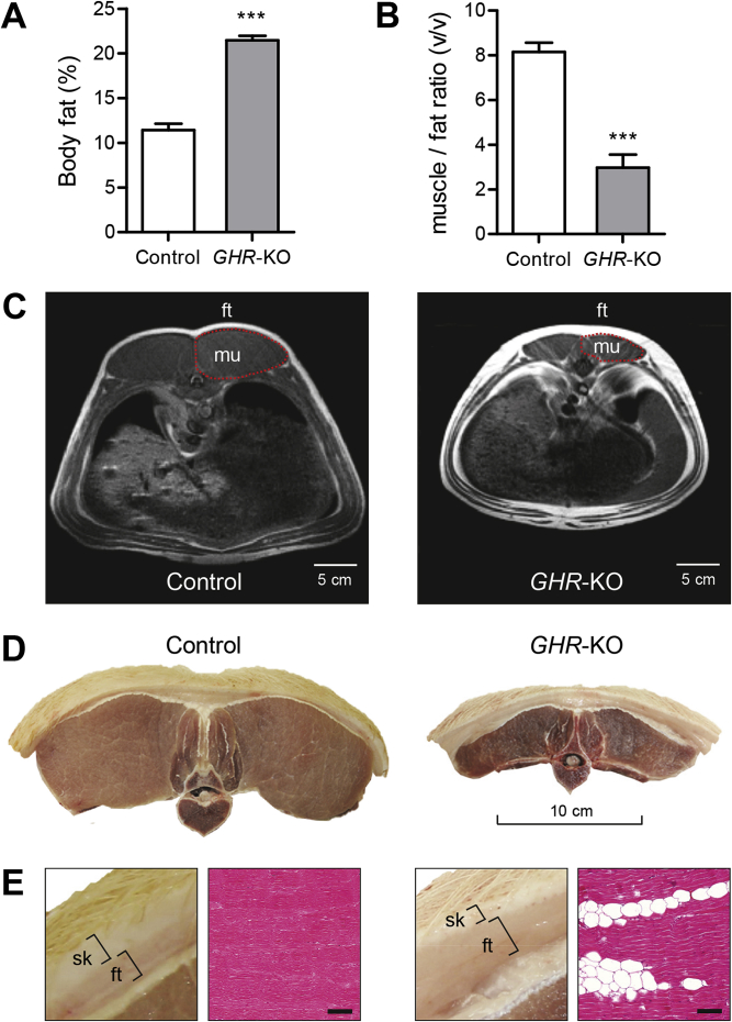
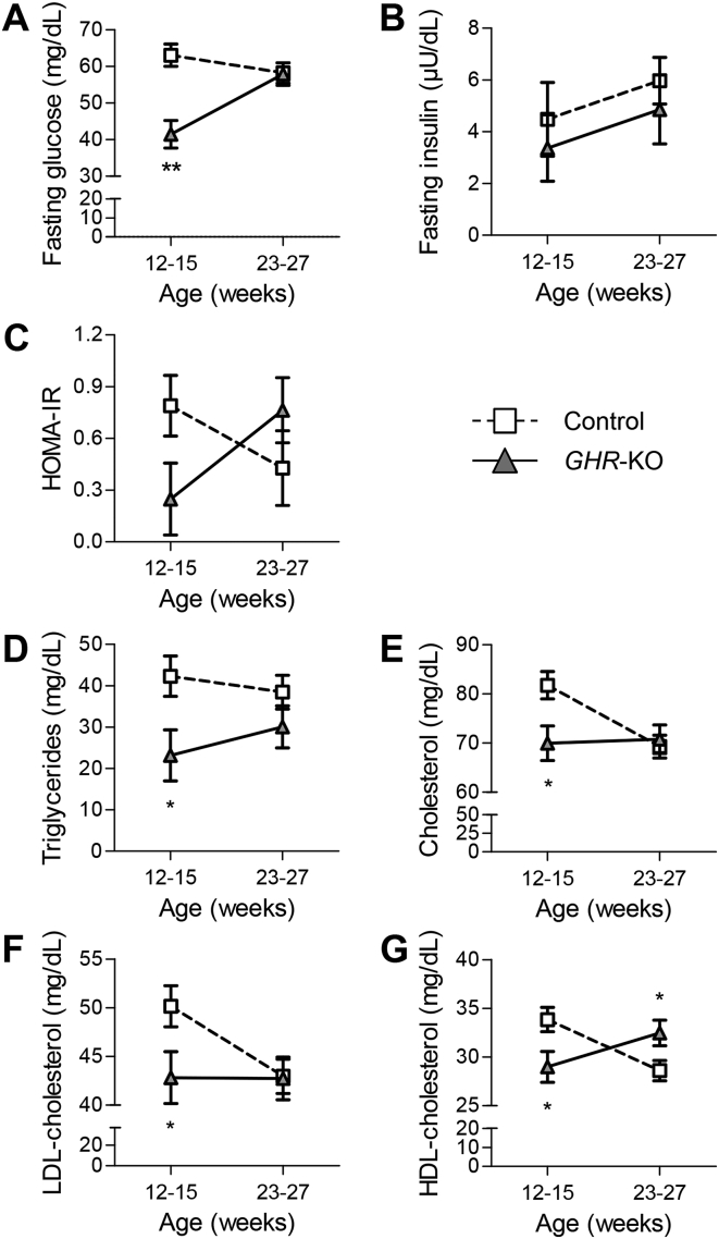
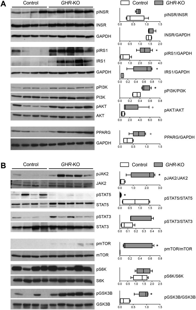
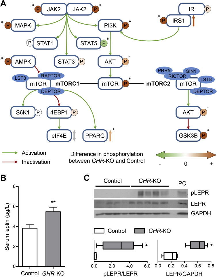
Results: GHR-KO pigs lacked GHR and had markedly reduced serum insulin-like growth factor 1 (IGF1) levels and reduced IGF-binding protein 3 (IGFBP3) activity but increased IGFBP2 levels. Serum GH concentrations were significantly elevated compared with control pigs. GHR-KO pigs had a normal birth weight. Growth retardation became significant at the age of five weeks. At the age of six months, the body weight of GHR-KO pigs was reduced by 60% compared with controls. Most organ weights of GHR-KO pigs were reduced proportionally to body weight. However, the weights of liver, kidneys, and heart were disproportionately reduced, while the relative brain weight was almost doubled. GHR-KO pigs had a markedly increased percentage of total body fat relative to body weight and displayed transient juvenile hypoglycemia along with decreased serum triglyceride and cholesterol levels. Analysis of insulin receptor related signaling in the liver of adult fasted pigs revealed increased phosphorylation of IRS1 and PI3K. In agreement with the loss of GHR, phosphorylation of STAT5 was significantly reduced. In contrast, phosphorylation of JAK2 was significantly increased, possibly due to the increased serum leptin levels and increased hepatic leptin receptor expression and activation in GHR-KO pigs. In addition, increased mTOR phosphorylation was observed in GHR-KO liver samples, and phosphorylation studies of downstream substrates suggested the activation of mainly mTOR complex 2.

Conclusion: GHR-KO pigs resemble the pathophysiology of LS and are an interesting model for mechanistic studies and treatment trials.

Keywords: Dwarfism; Growth hormone receptor; Hypoglycemia; Insulin-like growth factor 1; Laron syndrome; Pig model; Signaling.


Figures

Figure 1
Figure 1
Generation of a GHR-deficient pig model using CRISPR/Cas technology. (A) Partial DNA sequence of GHR exon 3. The sgRNA binding site is indicated in blue and the protospacer adjacent motif (PAM) in green. Insertions (red) of 1 bp (founder #2529) or 7 bp (founder #2533) lead to a shift of the reading frame. WT = wild type. (B) Restriction fragment length polymorphism analysis to detect the WT GHR sequence as well as monoallelic (Het) and biallelic (KO) mutations. (C) Partial amino acid sequences encoded by the WT and mutant GHR alleles. The signal peptide is shown in gray, WT GHR aa sequence in black (aa encoded by adjacent non-symmetrical exons in blue), missense aa sequence in red, and the premature termination codon as an asterisk. (D) Ligand immunohistochemistry demonstrating the absence of functional GHR (brown staining in control) in GHR-KO pigs. Chromogen: DAB; counterstain: Mayer's hemalum; bar = 10 μm.
Figure 2
Figure 2
Serum IGF1, IGFBP and GH concentrations of GHR-KO compared with control pigs. (A) Scatter plot of serum IGF1 levels of GHR-KO and control pigs over time. (B) Means and standard deviations of all serum IGF1 values displayed in panel A (GHR-KO: n = 42; control: n = 69). (C) Representative IGFBP ligand blot. Right lane displays recombinant human IGFBP3 (41/38 kDa), IGFBP2 (32 kDa), IGFBP5 (29 kDa) and IGFBP4 (24 kDa). (D) Quantification of IGFBP3 and IGFBP2 in serum from GHR-KO (n = 10) and control pigs (n = 12). The figure shows medians, 25th and 75th percentiles (box), and extremes (whiskers). (E) Representative GH secretion profiles of two female GHR-KO and two female control pigs. (F) Area under the GH curve (AUC; means and standard deviations for 6 female GHR-KO and 5 female/1 male control pigs). AU = arbitrary units. *p < 0.05; ***p < 0.001.
Figure 3
Figure 3
Body weight gain and growth of GHR-KO compared with control pigs. (A) GHR-KO pig (front) and control littermate aged 6 months. (B) Body weight gain. (C) Body length. (D) Relative body length (body length divided by the cube root of body weight). These parameters were determined in 12 GHR-KO and 25 control pigs. Panels A–D show least squares means (LSMs) and standard errors of LSMs estimated for group*age (see 2.8 for the statistical model). *p < 0.05; **p < 0.01; ***p < 0.001; ns = not significant.
Figure 4
Figure 4
Body composition of 6-month-old GHR-KO compared with control pigs. (A) DXA analysis revealed a significantly higher amount of total body fat in GHR-KO pigs. (B) The calculated ratio of muscle to fat tissue from MRI images at the last rib revealed a significant shift towards fat tissue in GHR-KO pigs (GHR-KO: n = 12; control: n = 25; ***p < 0.001). Panels A and B show least squares means (LSMs) and standard errors of LSMs estimated for the 2 groups (see 2.8 for the statistical model). (C) Representative magnetic resonance images used to evaluate the volume of the longissimus dorsi muscle (mu) and its overlying back fat (ft) at the last rib in GHR-KO and control pigs. Note the larger subcutaneous and visceral fat depots in GHR-KO pigs. (D) Representative macroscopic cross-sections of the first lumbar vertebra, the two longissimus dorsi muscles and the overlying back fat and skin. (E) Higher magnification of D showing an increased ratio of subcutaneous fat (ft) to skin (sk) thickness in a GHR-KO compared with a control pig. Histological section (H&E stain) showing an increased amount of intramuscular fat in GHR-KO pigs (bar = 100 μm).
Figure 5
Figure 5
Disproportionate organ growth in GHR-KO compared with control pigs. GHR-deficiency led to a proportionate and disproportionate reduction in organ sizes. (A) Representative organs from control (left) and GHR-KO pigs (right). (B) Relative differences between GHR-KO and control pigs in absolute organ weights and in organ weight-to-body weight ratios (relative organ weights). These parameters were determined in 9 GHR-KO and 25 control pigs, and least squares means (LSMs) and standard errors of LSMs were estimated for the 2 groups (see 2.8 for the statistical model). *p < 0.05; **p < 0.01; ***p < 0.001.
Figure 6
Figure 6
Age-dependent changes in glucose and lipid homeostasis parameters in GHR-KO and control pigs. (A) Transient juvenile hypoglycemia in GHR-KO pigs. (B) Unchanged serum insulin concentrations. (C) Initially lower, then higher HOMA-IR score (interaction group*age: p < 0.05). Serum concentrations of (D) triglycerides, (E) cholesterol, (F) low-density lipoprotein (LDL)-cholesterol, and (G) high-density lipoprotein (HDL)-cholesterol levels were significantly lower in young GHR-KO pigs than in age-matched controls, but normalized with age. HDL-cholesterol levels of 23- to 27-week-old GHR-KO pigs were even higher than in their control littermates. At least 6 animals per group and age-class were investigated. Panels A–G show least squares means (LSMs) and standard errors of LSMs estimated for group*age (see 2.8 for the statistical model). *p < 0.05; **p < 0.01.
Figure 7
Figure 7
Western blot analysis of signaling cascades in liver samples of 6-month-old fasted GHR-KO (n = 5) and control pigs (n = 4). (A) Insulin receptor-related signaling pathway and PPARG. (B) GHR- and mTOR-related signaling pathways. The box plots show medians, 25th and 75th percentiles (box), and extremes (whiskers). *p < 0.05; °p = 0.0635; evaluated using the Mann–Whitney U test.
Figure 8
Figure 8
(A) Schematic summary of the changes in phosphorylation in INSR- and GHR-related signaling molecules in liver samples of GHR-KO compared with control pigs. *p < 0.05; °p = 0.0635; evaluated using the Mann–Whitney U test. (B) Significantly increased fasting serum leptin concentrations in 6-month-old GHR-KO vs. control pigs. The figure shows the estimated least squares means (LSMs) and standard errors of the LSMs for the two groups, taking into account the effect of sex (9 male/13 female control pigs; 6 male/6 female GHR-KO pigs). **p < 0.01 for the effect of group (PROC GLM). (C) Significantly increased expression and phosphorylation of LEPR in liver samples from GHR-KO compared with control pigs. PC = protein lysate from choroid plexus of a wild-type pig used as positive control. *p < 0.05; evaluated using the Mann–Whitney U test.

Similar articles

Cited by

References

    1. Laron Z., Pertzelan A., Mannheimer S. Genetic pituitary dwarfism with high serum concentation of growth hormone–a new inborn error of metabolism? Israel Journal of Medical Sciences. 1966;2:152–155. - PubMed
    1. Laron Z. Clinical evidence of growth hormone resistance in patients with Laron syndrome. In: Laron Z., Kopchick J.J., editors. Laron Syndrome - from Man to Mouse. Springer; 2011. pp. 21–25.
    1. Laron Z. Laron syndrome (primary growth hormone resistance or insensitivity): the personal experience 1958–2003. The Journal of Clinical Endocrinology and Metabolism. 2004;89:1031–1044. - PubMed
    1. Laron Z. Epilogue: the future of Laron syndrome — the need for changes. Growth Hormone & IGF Research. 2016;28:79–80. - PubMed
    1. Guevara-Aguirre J., Rosenbloom A.L., Fielder P.J., Diamond F.B., Jr., Rosenfeld R.G. Growth hormone receptor deficiency in Ecuador: clinical and biochemical phenotype in two populations. The Journal of Clinical Endocrinology and Metabolism. 1993;76:417–423. - PubMed

Publication types

MeSH terms

Substances