. 2018 Jun;177(2):447-464.
doi: 10.1104/pp.18.00175. Epub 2018 Apr 20.

GSyellow, a Multifaceted Tag for Functional Protein Analysis in Monocot and Dicot Plants

Affiliations

GSyellow, a Multifaceted Tag for Functional Protein Analysis in Monocot and Dicot Plants

Nienke Besbrugge et al. Plant Physiol. 2018 Jun.

Abstract

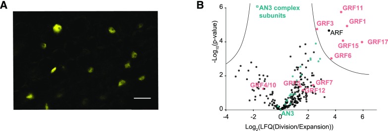
The ability to tag proteins has boosted the emergence of generic molecular methods for protein functional analysis. Fluorescent protein tags are used to visualize protein localization, and affinity tags enable the mapping of molecular interactions by, for example, tandem affinity purification or chromatin immunoprecipitation. To apply these widely used molecular techniques on a single transgenic plant line, we developed a multifunctional tandem affinity purification tag, named GSyellow, which combines the streptavidin-binding peptide tag with citrine yellow fluorescent protein. We demonstrated the versatility of the GSyellow tag in the dicot Arabidopsis (Arabidopsis thaliana) using a set of benchmark proteins. For proof of concept in monocots, we assessed the localization and dynamic interaction profile of the leaf growth regulator ANGUSTIFOLIA3 (AN3), fused to the GSyellow tag, along the growth zone of the maize (Zea mays) leaf. To further explore the function of ZmAN3, we mapped its DNA-binding landscape in the growth zone of the maize leaf through chromatin immunoprecipitation sequencing. Comparison with AN3 target genes mapped in the developing maize tassel or in Arabidopsis cell cultures revealed strong conservation of AN3 target genes between different maize tissues and across monocots and dicots, respectively. In conclusion, the GSyellow tag offers a powerful molecular tool for distinct types of protein functional analyses in dicots and monocots. As this approach involves transforming a single construct, it is likely to accelerate both basic and translational plant research.


Figures

Figure 1.
Figure 1.
Overview of the multifunctional GSyellow TAP tag molecular toolbox. Both C- and N-terminal fusions of the tag to the bait of interest are shown (middle). The GSyellow tag combines the cYFP with the SBP. Both tags are separated by tandem repeats of tobacco etch virus (TEV) and 3C rhinovirus (Rhino) protease cleavage sites, enabling gentle elution during TAP. The GSyellow tag allows protein complex analysis by AP-MS, either by TAP for highly specific isolation of clean protein complexes or by one-step anti-GFP-based immunoprecipitation (IP; top left). During TAP, complexes are isolated using anti-GFP immunoprecipitation, eluted with TEV or Rhino protease, and further purified using streptavidin beads. FN, False negatives reflecting bona fide interactions lost during AP-MS; FP, false positives isolated based on nonspecific interactions; TP, true positives representing bona fine bait interactions. Purified proteins are identified by MS. For analysis of protein-DNA interactions by ChIP-seq, anti-GFP-based ChIP protocols can be applied on a GSyellow transgenic line (top right). Purified DNA is analyzed by next generation sequencing to identify bound DNA fragments. Finally, the cYFP moiety of the GSyellow tag also enables subcellular localization analysis, both in Arabidopsis and maize (bottom left; bars = 50 μm). For targeting of TAP fusion proteins to specific organelles (bottom right), targeting peptide variants of both the N-terminal GSyellow and GSrhino TAP tags were developed, specifically targeting the nucleus, mitochondria, chloroplasts, or endoplasmic reticulum (ER), as shown by the organelle-targeting peptide variants of the GSrhino tag fused to GFP, transiently expressed in tobacco leaves (bars = 100, 50, 100, and 10 μm, respectively).
Figure 2.
Figure 2.
Functional analysis of GSyellow-tagged AN3 and TPLATE. A, Leaf areas of 21-d-old wild-type (WT) or AN3-GSyellow-overexpressing plants grown on control medium. The areas of cotyledons (cot) and leaves 1 to 9 (L1–L9) were measured from leaf series. Error bars represent se (n = 3), and asterisks indicate significant differences from the wild type (Col-0; P < 0.05, Student’s t test). B, Representative rosettes of 21-d-old plants used for leaf series. Bars = 1 cm. C, Confocal microscopy images of the root tip from a 6-d-old seedling expressing 35S:AN3-GSyellow, showing nuclear localization of AN3. Bar = 20 μm. D, Segregation analysis of the endogenous TPLATE locus in the offspring produced by selfing the heterozygous TPLATE mutant or the heterozygous TPLATE mutant complemented homozygously with pLat52:TPLATE-GSyellow. TT = TPLATE +/+, Tt = TPLATE +/−, tt = TPLATE −/−. χ2 values for the observed segregation ratios are indicated. The observed values do not differ significantly from the expected values for the indicated ratios (χ2 = 3.84 at P < 0.05). E, Scanning electron microscopy images of pollen grains derived from heterozygous TPLATE mutant plants, wild-type plants, and a complemented mutant heterozygous for the TPLATE mutation and homozygous for pLat52:TPLATE-GSyellow. Ratios of wild-type versus mutant (mut) pollen are indicated. Bars = 10 μm. F, Left, Representative spinning disc confocal microscope image of a hypocotyl epidermal cell belonging to a TPLATE mutant plant complemented with pLat52:TPLATE-GSyellow, taken from the corresponding movies. Bar = 5 μm. Middle, Discrete, dynamic foci represent the recruitment of TPLATE, which can be measured over time through a kymograph (arrowheads indicate the beginning and end of the signal). Right, TPLATE-GSyellow has an average dwell time of 20 s at the plasma membrane (n = 964).
Figure 3.
Figure 3.
Genome-wide ChIP-seq analysis of PPD2-GSyellow. ChIP was performed with an anti-GFP antibody on cell suspension cultures (PSB-D) transformed with 35S:PPD2-GSyellow. The data were normalized to a 35S:NLS-GSyellow-expressing PSB-D culture and compared with TChAP on PSB-D cultures transformed with 35S:PPD2-HBH (Gonzalez et al., 2015). A, Venn diagram of genes shared in each retrieved gene list after normalization. B, Genome-wide distribution of PPD2 DNA-binding sites in relation to the gene structure. Only peaks present in the intersects of both replicates were considered. UTR, Untranslated region. C, Distribution of the distance of peak summits to the nearest annotated translation start site (TLS) in bp. D, Graphic representation of the enriched G-box motif bound by PPD2, as determined through de novo motif analysis on the GSyellow ChIP data.
Figure 4.
Figure 4.
Functional analysis of ZmAN3-GSyellow along the maize leaf growth zone. A, Confocal microscopy image of the division zone of leaf 4 at 2 d after emerging, showing nuclear localization of ZmAN3. Bar = 20 µm. B, Relative intensity-based label-free quantification of copurified proteins identified with TAP on ZmAN3-GSyellow shown in a volcano plot. Comparison between the division zone and the expansion zone delineates significant changes of interaction partners between the zones. GRFs and AN3 core complex members are indicated in red and blue, respectively.
Figure 5.
Figure 5.
Identification of genome-wide ZmAN3-GSyellow binding sites through ChIP-seq on the growth zone of leaf 4 in maize. ChIP experiments were carried out in parallel on the full growth zone (cm 1–4) of leaf 4 at 2 d after emerging. A, Overview of the strategy used to identify genome-wide target genes of ZmAN3 through ChIP-seq. Normalization against two negative controls (NoAB and ZmNLS-GSyellow) was carried out on two ZmAN3 replicates. For each normalization method, peaks in common between the two replicates were identified if 50% or more of the peaks overlapped. To identify high-confidence peaks, only peaks were retained in the overlap of both normalization methods, and peaks locating in greater than 5-kb intergenic regions were removed. B, Distance distribution per negative control in bp of ZmAN3 peak summits to the closest translation start or stop site. C, Schematic representation of genome-wide ZmAN3-binding sites in relation to the gene structure. UTR, Untranslated region.
Figure 6.
Figure 6.
Analysis of ZmAN3-GSyellow leaf ChIP targets. A, The first Venn diagram represents the significant overlap of the ZmAN3-GSyellow maize leaf ChIP data with genes differentially expressed (DEG) in the tassel of the ZmAN3 mutant (hypergeometric test, P = 1.23E-50) or with genes identified by ChIP in the tassel using ZmAN3-GFP. In the second Venn diagram, the significant overlap (hypergeometric test, P = 1.53E-73) is shown between the leaf ZmAN3-GSyellow ChIP targets and the Arabidopsis cell culture TChAP targets, taking only genes into account for which an ortholog could be determined. ** = highly significant overlap. B, Sashimi plot obtained from the Integrative Genomics Viewer, demonstrating the binding of ZmAN3 to the promoter regions of ZmCycD7;1 (GRMZM2G058410) and TCP TF (GRMZM2G003944). The sequence reads of both ZmAN3 replicates and of the two control samples (ZmNLS and NoAB) are shown. The ZmCycD7;1 and TCP gene regions are represented by blue bars.

Similar articles

Cited by

References

    1. Blomme J, Van Aken O, Van Leene J, Jégu T, De Rycke R, De Bruyne M, Vercruysse J, Nolf J, Van Daele T, De Milde L, et al. (2017) The mitochondrial DNA-associated protein SWIB5 influences mtDNA architecture and homologous recombination. Plant Cell 29: 1137–1156 - PMC - PubMed
    1. Bunnik EM, Le Roch KG (2013) An introduction to functional genomics and systems biology. Adv Wound Care (New Rochelle) 2: 490–498 - PMC - PubMed
    1. Bürckstümmer T, Bennett KL, Preradovic A, Schütze G, Hantschel O, Superti-Furga G, Bauch A (2006) An efficient tandem affinity purification procedure for interaction proteomics in mammalian cells. Nat Methods 3: 1013–1019 - PubMed
    1. Cheeseman IM, Desai A (2005) A combined approach for the localization and tandem affinity purification of protein complexes from metazoans. Sci STKE 2005: pl1. - PubMed
    1. Coussens G, Aesaert S, Verelst W, Demeulenaere M, De Buck S, Njuguna E, Inzé D, Van Lijsebettens M (2012) Brachypodium distachyon promoters as efficient building blocks for transgenic research in maize. J Exp Bot 63: 4263–4273 - PubMed

Publication types

MeSH terms