. 2018 May 16;38(20):4695-4707.
doi: 10.1523/JNEUROSCI.2951-17.2018. Epub 2018 Apr 20.

Distribution of Spinal Neuronal Networks Controlling Forward and Backward Locomotion

Affiliations

Distribution of Spinal Neuronal Networks Controlling Forward and Backward Locomotion

Natalia Merkulyeva et al. J Neurosci. .

Abstract

Higher vertebrates, including humans, are capable not only of forward (FW) locomotion but also of walking in other directions relative to the body axis [backward (BW), sideways, etc.]. Although the neural mechanisms responsible for controlling FW locomotion have been studied in considerable detail, the mechanisms controlling steps in other directions are mostly unknown. The aim of the present study was to investigate the distribution of spinal neuronal networks controlling FW and BW locomotion. First, we applied electrical epidural stimulation (ES) to different segments of the spinal cord from L2 to S2 to reveal zones triggering FW and BW locomotion in decerebrate cats of either sex. Second, to determine the location of spinal neurons activated during FW and BW locomotion, we used c-Fos immunostaining. We found that the neuronal networks responsible for FW locomotion were distributed broadly in the lumbosacral spinal cord and could be activated by ES of any segment from L3 to S2. By contrast, networks generating BW locomotion were activated by ES of a limited zone from the caudal part of L5 to the caudal part of L7. In the intermediate part of the gray matter within this zone, a significantly higher number of c-Fos-positive interneurons was revealed in BW-stepping cats compared with FW-stepping cats. We suggest that this region of the spinal cord contains the network that determines the BW direction of locomotion.SIGNIFICANCE STATEMENT Sequential and single steps in various directions relative to the body axis [forward (FW), backward (BW), sideways, etc.] are used during locomotion and to correct for perturbations, respectively. The mechanisms controlling step direction are unknown. In the present study, for the first time we compared the distributions of spinal neuronal networks controlling FW and BW locomotion. Using a marker to visualize active neurons, we demonstrated that in the intermediate part of the gray matter within L6 and L7 spinal segments, significantly more neurons were activated during BW locomotion than during FW locomotion. We suggest that the network determining the BW direction of stepping is located in this area.

Keywords: backward and forward walking; c-Fos; decerebrate cat; locomotor networks.


Figures

Figure 1.
Figure 1.
Experimental design. A, The head, vertebral column and pelvis of a decerebrate cat were fixed in a rigid frame (points of fixation are indicated by X). The hindlimbs were positioned on the treadmill. Walking of the hindlimbs was evoked by electrical ES of the spinal cord. A–P movements of each limb were recorded by a sensor (only the right sensor, Limb-R, is shown). To evoke FW and BW locomotion, during ES, the treadmill belt was moved backward and forward, respectively, in relation to the cat. Red circles are reflective markers attached to the skin projections of the main hindlimb joints. B, The side view of the walking cat was recorded with a video camera (Video). C, Examples of FW and BW locomotion evoked by ES of the L5 segment. Limb-R and Limb-L, movements of the right and left hindlimbs, respectively. EMGs from right (R) and left (L) TA and LG. HL(R) and HL(L), structure of locomotor cycles of the right and left limbs, respectively. D, Hindlimb configuration at different moments of the swing and stance during FW and BW locomotion. E1, E2, E3, and F: end of the swing–beginning of stance, stance, end of the stance–beginning of the swing and swing, respectively. Configurations corresponding to the shortest limb length during the swing and the longest limb length during the stance (at E3) are indicated in red. Black horizontal arrows indicate the direction of the limb movement during the stance and swing of FW and BW locomotion.
Figure 2.
Figure 2.
c-Fos Labeling of spinal neurons of locomotor networks and analysis of distribution of c-Fos-positive neurons. A, Sites of ES application for initiation of BW and FW locomotion in individual cats used for c-Fos labeling of spinal neurons. Fw1, Fw2, Fw3 and Bw1, Bw2, Bw3 are cats that performed FW and BW locomotion, respectively. B, Distribution of FOS+ nuclei on a frontal section of L7 in the cat Fw2. Borders between Rexed laminae are indicated by interrupted lines. Rexed laminae were segregated on the basis of morphological criteria described by Rexed (1954). Right, Magnifications of the red boxed areas containing labeled interneuronal nuclei (box 1), motoneuronal nuclei marked by black arrows (box 2) and unlabeled motoneuronal nuclei marked by white arrows (box 3). Scale bar, 200 μm. C, Procedure for visualizing the density of the FOS+ nuclei distributed over the cross section of a particular spinal segment (see Material and Methods). Bottom left, A color map of the density of FOS+ nuclei.
Figure 3.
Figure 3.
Efficacy of ES of different lumbosacral segments to evoke FW and BW locomotion. A, Effects of ES observed in individual animals. Each horizontal line shows the results obtained in an individual animal. Thick lines indicate segments (or parts of the segments) subjected to ES, and their color encodes the effect of stimulation (red, FW stepping; blue, BW stepping; pink, absence of FW stepping; light green, absence of BW stepping). B, Proportion of cats in which ES of a particular segment (or a part of the segment) evoked FW (red line) and BW (blue line) locomotion. C, The mean value of the optimal strength of ES for cats subjected to ES of a particular segment (or a part of the segment) is shown by the red line. The SE is indicated by the pink shading. The maximum strength of ES applied to a particular segment in an attempt to initiate FW or BW stepping is shown by a gray line.
Figure 4.
Figure 4.
Comparison of kinematic characteristics of FW stepping movements evoked by ES of different lumbosacral segments and BW stepping movements. A, Angles of the hindlimb joints at maximal limb flexion during swing (Swing angle) and maximal limb extension during stance (Stance angle). B, Step length. C, The range of hindlimb joint angles changes during the locomotor cycle (Stance angle − Swing angle, the difference between Stance angle and Swing angle averaged over all hindlimb joints in all cats). D, Stability of the limb locomotor pattern (self-similarity coefficient). E, Left-right step length asymmetry (coefficient of asymmetry). Kinematic characteristics were averaged over 120 FW locomotor cycles in four cats and over 45 BW locomotor cycles in three cats (mean ± SE; indication of significance level: *p < 0.05, ***p < 0.001).
Figure 5.
Figure 5.
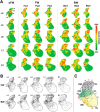
Distribution of FOS+ nuclei across the gray matter in segments L4–S1 of cats that performed FW and BW locomotion. A, Density of the FOS+ nuclei distribution across the left half of the gray matter in segments L4–S1 of individual cats. In color-coded images, the green-to-red gradient corresponds to the optical density of the gray level image's loci after the Gaussian blur. This gradient is individual for each segment in each cat. B, Distribution of FOS+ nuclei across the gray matter in segments L4–S1 of all cats that performed FW and BW locomotion. For each segment, the image shows the location of all FOS+ nuclei in the left half of gray matter combined from 15 sections taken from three cats (5 sections from each cat). FW and BW, cats that performed FW and BW locomotion, respectively, for 1.5–2 h. sFW, the cat that performed FW locomotion for 30 min. C, Subdivision of the gray matter into six areas (modified from Matsushita, 1970; see text for explanation). DL, Light gray; DM, dark gray; CL, light green; CM, dark green; VL, light yellow; VM, dark yellow.
Figure 6.
Figure 6.
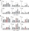
Comparison of the distribution of FOS+ nuclei in cats that performed FW and BW stepping. A, Comparison of the number of FOS+ nuclei in each of 6 areas (DL, DM, CL, CM, VL, and VM) of the gray matter in segments L4–S1 of cats that performed FW and BW stepping. The average number (±SE) of FOS+ nuclei revealed in 15 sections taken from three cats (5 sections from each cat) is presented for each area of a particular segment. B, Comparison of a number of FOS+ nuclei in each of six areas (DL, DM, CL, CM, VL, and VM) of the gray matter in segments L4–S1 of Fw2 and Bw2. Forward and backward locomotion was evoked in Fw2 and Bw2, respectively, by ES of L6 (marked by red). Indication of significance level: *p < 0.05, **p < 0.01.
Figure 7.
Figure 7.
Specificity of c-Fos immunostaining. A, The mean numbers (±SE) of FOS+ nuclei in the DL, DM, CL, CM, VL, and VM areas of the gray matter in segments L4–S1 of the cat (sFW) that performed a short period of stepping (30 min) and in cats (FW) that performed long-lasting stepping (1.5–2 h). For the cat sFW, an average based on five sections from each segment is presented. For FW cats, an average based on 15 sections from each segment taken from three cats (5 sections from each segment of each individual cat) is presented. BE, Kinematics of locomotor movements (B, C) and distribution of FOS+ nuclei (D, E) in two cats that performed symmetrical and asymmetrical walking (Bw2 and Bw1, respectively). B, Recording of locomotor movements of the right and left limbs (Limb-R and Limb-L) in Bw2 and Bw1. sw, Swing; st, stance; fl, a part of the locomotor cycle when the flexed limb is maintained above the surface at the extreme posterior position (relative to the trunk) before landing. C, Mean (±SE) of the step length (n = 50) exhibited by the left (L; light gray) and right (R; dark gray) hindlimb of Bw2 and Bw1. D, The coefficient of asymmetry (mean±SE) between the left and right hindlimbs in Bw2 and Bw1. E, The number of FOS+ nuclei on the left (L; light gray) and right (R; dark gray) sides of segments L4–S1 revealed in Bw2 and Bw1. The total number of FOS+ nuclei in five sections from each segment is shown.
Figure 8.
Figure 8.
Distribution of spinal networks generating FW and BW stepping. A, A hypothesis about the control of step direction (modified from Musienko et al., 2012). The locomotor system includes two principal mechanisms, one generating a vertical component of the step (limb elevation and lowering) and the other generating a horizontal component (limb transfer from one extreme point to the other). The latter includes networks generating the horizontal component of step in different directions (for simplicity, only the networks generating steps in four directions are shown: F, forward; B, backward; R, rightward; and L, leftward). These networks receive sensory input signaling limb motion in stance; reaching an extreme position triggers a limb transfer. ES of the spinal cord activates a network generating a vertical component of the step. It also causes subthreshold activation of all networks generating a horizontal component. Due to the treadmill motion (e.g., forward), the limb will reach an extreme anterior position, and sensory input will activate network B (marked in blue), which will evoke the BW step. Thus, ES stimulation can evoke stepping opposite to the direction of treadmill motion. B, Areas of the gray matter in L6 and L7 where the neurons of the network generating the horizontal component of BW steps are located (marked in blue). Abbreviations are as in Figure 5C. C, A scheme for the rostrocaudal distribution of a network generating the vertical component of the step (green thick line), a network generating the horizontal component for FW steps (red thick line) and a network generating the horizontal component for BW steps (blue thick line) in the lumbosacral enlargement. Insets 1–4, Hindlimb configurations in the middle of swing (pink and light blue) and at extreme limb positions during one step cycle (red and blue). Thick and thin black arrows show the direction of the treadmill motion and the directions of the limb swing movement, respectively. ES of rostral (L4–L5) and caudal (S1–S2) segments containing only two of three networks (generating the vertical component of the step and the horizontal component for the FW step) evokes FW locomotion (insets 1 and 4). ES of L6–L7 segments containing all three networks (generating the vertical component of the step, the horizontal component for the FW step and the horizontal component for the BW step), depending on the direction of the treadmill belt motion, evokes FW (inset 2) or BW (inset 3) stepping, respectively. Note that FW-stepping movements evoked by ES of rostral segments are performed at a more rostral position in relation to the trunk and with more flexed limb compared with those evoked by ES of caudal segments (compare insets 1 and 4).

Similar articles

Cited by

References

    1. Ahn SN, Guu JJ, Tobin AJ, Edgerton VR, Tillakaratne NJ (2006) Use of c-fos to identify activity-dependent spinal neurons after stepping in intact adult rats. Spinal Cord 44:547–559. 10.1038/sj.sc.3101862 - DOI - PMC - PubMed
    1. Arshavsky YI, Berkinblit MB, Gelfand IM, Orlovsky GN, Fukson OI (1972a) Recordings of neurons of the dorsal spinocerebellar tract during evoked locomotion. Brain Res 43:272–275. 10.1016/0006-8993(72)90295-8 - DOI - PubMed
    1. Arshavsky YI, Berkinblit MB, Gelfand IM, Orlovsky GN, Fukson OI (1972b) Origin of modulation of the ventral spinocerebellar tract during locomotion. Brain Res 43:276–279. 10.1016/0006-8993(72)90296-X - DOI - PubMed
    1. Arshavsky YI, Gelfand IM, Orlovsky GN (1986) Cerebellum and rhythmical movements. New York: Springer.
    1. Barthélemy D, Leblond H, Rossignol S (2007) Characteristics of mechanisms of locomotion induced by intraspinal microstimulation and dorsal root stimulation in spinal cats. J Neurophysiol 97:1986–2000. 10.1152/jn.00818.2006 - DOI - PubMed

Publication types

Substances

LinkOut - more resources