. 2018 Jun 29;293(26):9937-9944.
doi: 10.1074/jbc.RA118.002494. Epub 2018 Apr 20.

Potassium is a trigger for conformational change in the fusion spike of an enveloped RNA virus

Affiliations

Potassium is a trigger for conformational change in the fusion spike of an enveloped RNA virus

Emma K Punch et al. J Biol Chem. .

Erratum in

Abstract

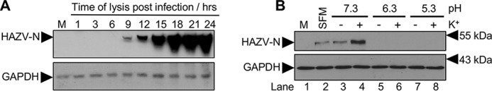
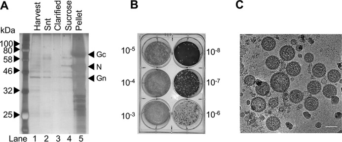
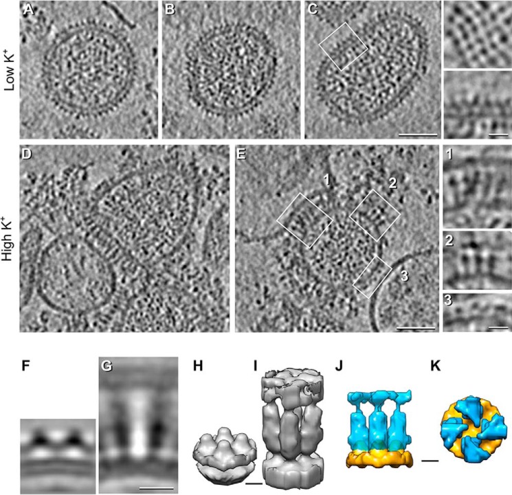
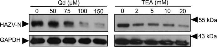
Many enveloped viruses enter cells through the endocytic network, from which they must subsequently escape through fusion of viral and endosomal membranes. This membrane fusion is mediated by virus-encoded spikes that respond to the dynamic endosomal environment, which triggers conformational changes in the spikes that initiate the fusion process. Several fusion triggers have been identified and include pH, membrane composition, and endosome-resident proteins, and these cues dictate when and where viral fusion occurs. We recently reported that infection with an enveloped bunyavirus requires elevated potassium ion concentrations [K+], controlled by cellular K+ channels, that are encountered during viral transit through maturing endosomes. Here we reveal the molecular basis for the K+ requirement of bunyaviruses through the first direct visualization of a member of the Nairoviridae family, namely Hazara virus (HAZV), using cryo-EM. Using cryo-electron tomography, we observed HAZV spike glycoproteins within infectious HAZV particles exposed to both high and low [K+], which showed that exposure to K+ alone results in dramatic changes to the ultrastructural architecture of the virion surface. In low [K+], the spikes adopted a compact conformation arranged in locally ordered arrays, whereas, following exposure to high [K+], the spikes became extended, and spike-membrane interactions were observed. Viruses exposed to high [K+] also displayed enhanced infectivity, thus identifying K+ as a newly defined trigger that helps promote viral infection. Finally, we confirmed that K+ channel blockers are inhibitory to HAZV infection, highlighting the potential of K+ channels as anti-bunyavirus targets.

Keywords: conformational change; electron tomography; fusion protein; ion channel; potassium transport; virus entry; virus structure.


Conflict of interest statement

The authors declare that they have no conflicts of interest with the contents of this article

Figures

Figure 1.
Figure 1.
HAZV infectivity can be increased by exposure to elevated [K+]. A, time course of HAZV multiplication in A549 cells infected at an m.o.i. of 0.1 or mock-infected (M) as assessed by HAZV N protein production, detected by Western blotting with anti-HAZV N antiserum alongside GAPDH loading controls. Lanes are labeled with time of harvest after infection in hours. A long exposure of the Western blotting is shown to reveal early N production. B, HAZV was exposed in vitro to either low (5 mm, −K+) or high (140 mm, +K+) relative [K+] at a pH of 5.3, 6.3, or 7.3. After incubation, [K+] were diluted by addition of SFM (K+ concentration, 5.3 mm), and K+-exposed virions were used to infect A549 cells at an m.o.i. of 0.1 for 18 h. Cells were also mock-infected and incubated with virus incubated in SFM alone. The abundance of HAZV N protein expression was assessed by Western blot analysis as described for A, with a representative blot shown here.
Figure 2.
Figure 2.
Purification and cryo-EM of HAZV particles. A, HAZV was harvested from infected cell supernatant (Snt), which was clarified and then pelleted by centrifugation through a sucrose cushion, and analyzed by SDS-PAGE. B, resuspended HAZV was titered on SW13 cells. C, HAZV was vitrified on carbon-backed grids for cryo-EM analysis, which revealed a pleomorphic virion morphology, with viral glycoprotein spikes visible around the perimeter. Scale bar = 100 nm.
Figure 3.
Figure 3.
Cryo-ET and STA of HAZV virions treated with low or high [K+] at pH 7.3. A–E, central tomographic sections of HAZV virions. A–C, low [K+] (5 mm). A continuous glycoprotein array around the viral envelope is evident. The 4-fold arrangement of the spikes is shown in a tangential section of a HAZV virion (C, top inset). Magnification of the squared region in C is shown in the bottom inset. D and E, high [K+] (140 mm). Changes in [K+] resulted in extension of the glycoprotein spikes (e.g. E, insets 1 and 2) and interactions with adjacent membranes (e.g. E, inset 1) co-purified with HAZV virions. E, inset 3, shows control-like spikes in high [K+]–treated HAZV virions. F–K, STA of HAZV spikes: Sagittal sections (F and G) and isosurface (H and I) rendering of HAZV spikes at low (F and H) and high (G and I) [K+]. J and K, superimposed isosurface rendering of low (orange) and high (blue) [K+]–treated segmented spikes as seen from the side (J) and top (K). Although the high K+ average is ∼3 times longer than the low K+ average (J), the low K+ average shows a continuous density parallel to the viral envelope that is absent in the high K+ average (K). Scale bars = 50 nm in A–E, 10 nm in F and G, and 5 nm in H–K.
Figure 4.
Figure 4.
Model describing the HAZV glycoprotein rearrangements after exposure to high K+.
Figure 5.
Figure 5.
HAZV multiplication can be blocked by broad-spectrum inhibition of cellular K+ channels. Representative western blots of A549 lysates following pretreatment with the indicated channel blockers (TEA, and Qd) prior to infection with HAZV (m.o.i. of 0.1). After 24 h, cell lysates were probed by Western blotting with sheep anti-HAZV N serum and GAPDH as a loading control. No-treatment controls were included for each inhibitor.

Similar articles

Cited by

References

    1. Walter C. T., and Barr J. N. (2011) Recent advances in the molecular and cellular biology of bunyaviruses. J. Gen. Virol. 92, 2467–2484 10.1099/vir.0.035105-0 - DOI - PubMed
    1. Adams M. J., Lefkowitz E. J., King A. M. Q., Harrach B., Harrison R. L., Knowles N. J., Kropinski A. M., Krupovic M., Kuhn J. H., Mushegian A. R., Nibert M., Sabanadzovic S., Sanfaçon H., Siddell S. G., Simmonds P., et al. (2017) Changes to taxonomy and the International Code of Virus Classification and Nomenclature ratified by the International Committee on Taxonomy of Viruses (2017). Arch. Virol. 162, 2505–2538 10.1007/s00705-017-3358-5 - DOI - PubMed
    1. Estrada-Peña A., Ruiz-Fons F., Acevedo P., Gortazar C., and de la Fuente J. (2013) Factors driving the circulation and possible expansion of Crimean-Congo haemorrhagic fever virus in the western Palearctic. J. Appl. Microbiol. 114, 278–286 10.1111/jam.12039 - DOI - PubMed
    1. Suleiman M. N., Muscat-Baron J. M., Harries J. R., Satti A. G., Platt G. S., Bowen E. T., and Simpson D. I. (1980) Congo/Crimean haemorrhagic fever in Dubai: an outbreak at the Rashid Hospital. Lancet 2, 939–941 - PubMed
    1. Surtees R., Ariza A., Punch E. K., Trinh C. H., Dowall S. D., Hewson R., Hiscox J. A., Barr J. N., and Edwards T. A. (2015) The crystal structure of the Hazara virus nucleocapsid protein. BMC Struct. Biol. 15, 24 10.1186/s12900-015-0051-3 - DOI - PMC - PubMed

Publication types

MeSH terms