. 2018 Jul;24(7):939-949.
doi: 10.1261/rna.066415.118. Epub 2018 Apr 20.

Role of the novel endoribonuclease SLFN14 and its disease-causing mutations in ribosomal degradation

Affiliations

Role of the novel endoribonuclease SLFN14 and its disease-causing mutations in ribosomal degradation

Sarah J Fletcher et al. RNA. 2018 Jul.

Abstract

Platelets are anucleate and mostly ribosome-free cells within the bloodstream, derived from megakaryocytes within bone marrow and crucial for cessation of bleeding at sites of injury. Inherited thrombocytopenias are a group of disorders characterized by a low platelet count and are frequently associated with excessive bleeding. SLFN14 is one of the most recently discovered genes linked to inherited thrombocytopenia where several heterozygous missense mutations in SLFN14 were identified to cause defective megakaryocyte maturation and platelet dysfunction. Yet, SLFN14 was recently described as a ribosome-associated protein resulting in rRNA and ribosome-bound mRNA degradation in rabbit reticulocytes. To unveil the cellular function of SLFN14 and the link between SLFN14 and thrombocytopenia, we examined SLFN14 (WT/mutants) in in vitro models. Here, we show that all SLFN14 variants colocalize with ribosomes and mediate rRNA endonucleolytic degradation. Compared to SLFN14 WT, expression of mutants is dramatically reduced as a result of post-translational degradation due to partial misfolding of the protein. Moreover, all SLFN14 variants tend to form oligomers. These findings could explain the dominant negative effect of heterozygous mutation on SLFN14 expression in patients' platelets. Overall, we suggest that SLFN14 could be involved in ribosome degradation during platelet formation and maturation.

Keywords: RNA degradation; SLFN14; endoribonuclease; platelet; ribonuclease; thrombocytopenia.


Figures

FIGURE 1.
FIGURE 1.
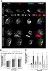
Significant colocalization is observed between SLFN14 (WT/mut)-myc and 5.8S rRNA in differentiated Dami cells. There is no alteration in subcellular distribution between SLFN14 (WT)-myc and SLFN14 (mut)-myc distribution or colocalization with 5.8S rRNA. (A) Transiently transfected differentiated Dami cells expressing SLFN14(WT/mut)-myc for 48 h were probed with rabbit anti-myc and mouse anti-5.8S primary antibodies followed by incubation with anti-rabbit AlexaFluor488, anti-mouse AlexaFluor568 secondary antibodies and TO-PRO-3 Iodide nuclear stain. A representative image is shown from three independent experiments, scale bar denotes 15 µm. (B) Pearson's correlation coefficient data demonstrating no change in colocalization or subcellular-distribution between 5.8S rRNA and SLFN14 (WT)-myc or SLFN14 (mut)-myc in comparison to control areas. n = at least 40 cells analyzed from three independent experiments. (C) Pearson's correlation coefficient data demonstrating a significant increase in colocalization between 5.8S rRNA and SLFN14 (WT/mut)-myc staining in comparison to control areas. (***) P ≤ 0.001 colocalization between 5.8S rRNA and SLFN14 (WT/mut)-myc and control area. Error bars ± SD.
FIGURE 2.
FIGURE 2.
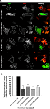
Reduced intensity of 5.8S rRNA staining in differentiated Dami cells expressing wild-type/mutant SLFN14 constructs. (A) Transiently transfected Dami cells expressing SLFN14(WT/mut)-myc for 48 h were probed with rabbit anti-myc and mouse anti-5.8S primary antibodies followed by incubation with anti-rabbit AlexaFluor488 and anti-mouse AlexaFluor568 secondary antibodies, respectively. The dashed white line outlines cells expressing SLFN14 (WT/mut)-myc, the solid line represents the outline of cells which were not transfected. A representative image is shown from three independent experiments. Scale bar denotes 15 µm. (B) Average intensity measurements from the entire cell area were quantified from images represented in A. n = at least 40 cells analyzed from three independent experiments. (***) P ≤ 0.001 when compared to nontransfected cells. Error bars ± SD.
FIGURE 3.
FIGURE 3.
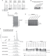
Association of SLFN14 (WT/mut)-myc with ribosomes and ribosomal subunits resulting in rRNA endonucleolytic degradation in HEK293T cells. (A) Binding of SLFN14 (WT/mut)-myc to 80S monosomes, 60S, and 40S ribosomal subunits obtained after SDG centrifugation of HEK293T cell lysates with overexpressed one of SLFN14 (WT/mut)-myc forms. Assignment of 80S monosomes, 40S, and 60S subunits in gradient fractions was based on immunoblotting with anti-RPS19 and anti-RPL3 antibodies as exemplified by the EV control. Ribosomal fractions were concentrated and analyzed by immunoblotting with anti-SLFN14 antibodies. (B) Expression levels of SLFN14 (WT/mut)-myc in HEK293T cells assayed by immunoblotting with anti-SLFN14 and anti-GAPDH (control) antibodies. (C) Binding of recombinant SLFN14 (WT)-65 kDa and SLFN14 (K218E)-65 kDa to assembled 80S ribosomes assayed by SDG centrifugation and Coomassie staining. (D) rRNA degradation in SLFN14(WT/mut)-overexpressed HEK293T cells assayed by denaturing agarose/formaldehyde gel electrophoresis (n = 3 independent experiments). The asterisks indicate the main bands of rRNA fragments. Positions of the 28S rRNA and 18S rRNA are shown. (E) Integrity analysis of rRNA. (Left panel) Representative electrophoretic profiles of total RNA obtained using the Agilent 2200 TapeStation system from SLFN14(WT/mut)-overexpressed HEK293T cells. Arrows indicate the appearance of rRNA degradation fragments. (Right panel) representative data output gel-like images. The average RIN scores from three independent experiments are shown.
FIGURE 4.
FIGURE 4.
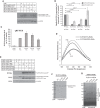
Role of SLFN14 missense mutations in protein expression. (A) Immunoblotting image showing levels of SLFN14 (WT/mut)-myc and SLFN14 (WT)-GFP in HEK293T cells transiently expressing the above constructs. The blot was probed with anti-SLFN14 and anti-GAPDH primary antibodies followed by incubation with anti-rabbit HRP. (B) Quantification of SLFN14 (WT/mut)-myc and SLFN14 (WT)-GFP protein expression from immunoblotting analysis of n = 3 lysate samples per condition from three independent experiments. All values are mean ± SD. (C) Quantification of relative SLFN14 (WT/mut)-myc transcript level normalized to GAPDH control transcript from qRT-PCR test data on n = 6 independent sets of lysate samples. All values are mean ± SD. (D) Fluorescence emission spectra of SLFN14 (WT)-45 kDa and SLFN14 (K218E)-45 kDa proteins collected at the same protein concentration at two different temperatures: 25°C and 65°C (n = 3 independent experiments). (E) Coimmunoprecipitation (co-IP) analysis in HEK293T cells to assess both wild-type and mutant GFP labeled SLFN14 with SLFN14(WT)-myc to look at formation of hetero-oligomers between wild-type and mutant SLFN14. (F) Purified recombinant SLFN14-45 kDa WT and mutants resolved by SDS-PAGE. (G) Oligomerization capacity of SLFN14-45 kDa WT and mutants assayed by native PAGE. Positions of different oligomeric forms are shown.

References

    1. Angénieux C, Maître B, Eckly A, Lanza F, Gachet C, de la Salle H. 2016. Time-dependent decay of mRNA and ribosomal RNA during platelet aging and its correlation with translation activity. PLoS One 11: e0148064. - PMC - PubMed
    1. Berger M, Krebs P, Crozat K, Li X, Croker BA, Siggs OM, Popkin D, Du X, Lawson BR, Theofilopoulos AN, et al. 2010. An Slfn2 mutation causes lymphoid and myeloid immunodeficiency due to loss of immune cell quiescence. Nat Immunol 11: 335–343. - PMC - PubMed
    1. Brady G, Boggan L, Bowie A, O'Neill LAJ. 2005. Schlafen-1 causes a cell cycle arrest by inhibiting induction of cyclin D1. J Biol Chem 280: 30723–30734. - PubMed
    1. Cox TM, Cachón-González MB. 2012. The cellular pathology of lysosomal diseases. J Pathol 226: 241–254. - PubMed
    1. Favier R, Raslova H. 2015. Progress in understanding the diagnosis and molecular genetics of macrothrombocytopenias. Br J Haematol 170: 626–639. - PubMed

Publication types

LinkOut - more resources