. 2018 Apr 20;9(1):1583.
doi: 10.1038/s41467-018-03926-1.

Self-replication of DNA by its encoded proteins in liposome-based synthetic cells

Affiliations

Self-replication of DNA by its encoded proteins in liposome-based synthetic cells

Pauline van Nies et al. Nat Commun. .

Abstract

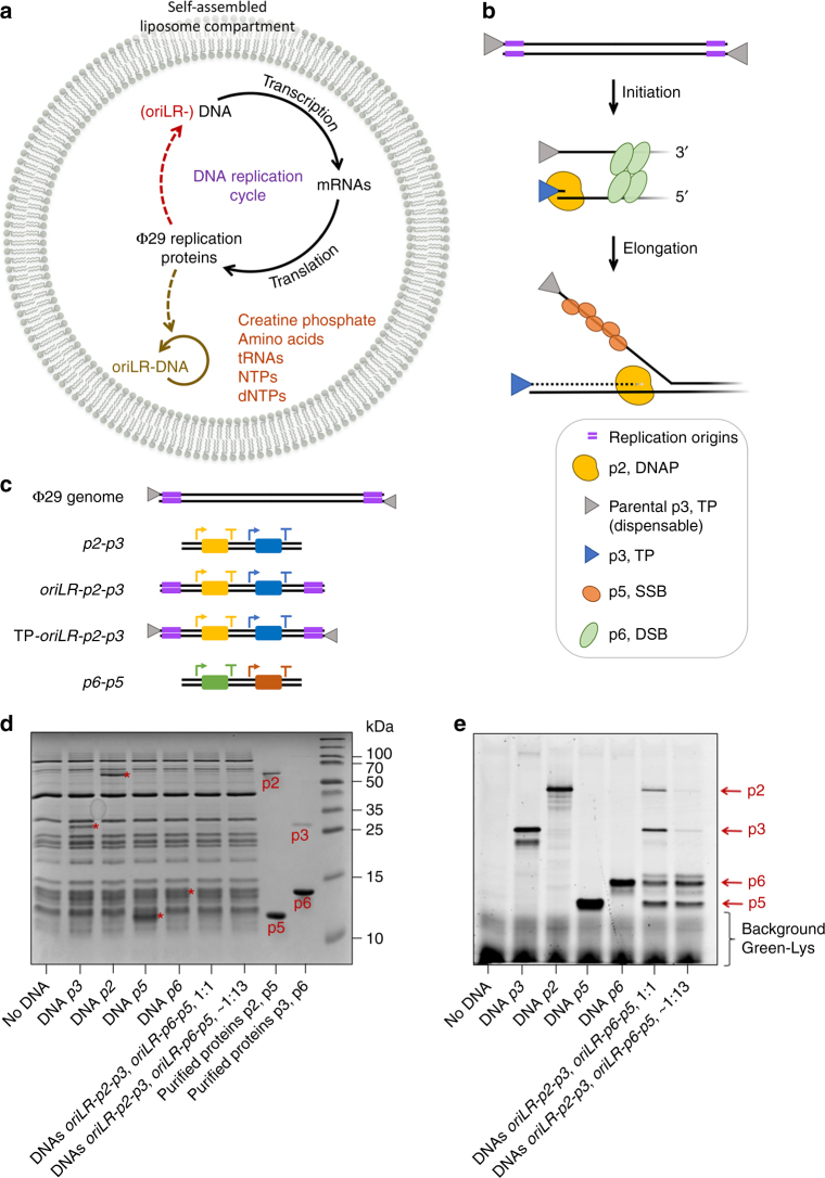
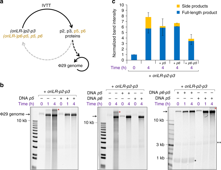
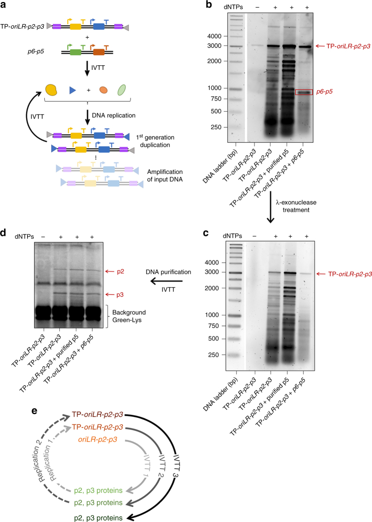
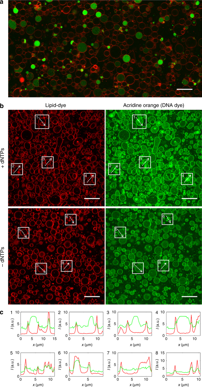
Replication of DNA-encoded information and its conversion into functional proteins are universal properties of life. In an effort toward the construction of a synthetic minimal cell, we implement here the DNA replication machinery of the Φ29 virus in a cell-free gene expression system. Amplification of a linear DNA template by self-encoded, de novo synthesized Φ29 proteins is demonstrated. Complete information transfer is confirmed as the copied DNA can serve as a functional template for gene expression, which can be seen as an autocatalytic DNA replication cycle. These results show how the central dogma of molecular biology can be reconstituted and form a cycle in vitro. Finally, coupled DNA replication and gene expression is compartmentalized inside phospholipid vesicles providing the chassis for evolving functions in a prospective synthetic cell relying on the extant biology.


Conflict of interest statement

The authors declare no competing interests.

Figures

Fig. 1
Fig. 1
Basic elements of the DNA self-replication strategy. a Flow of genetic information reconstituted in batch mode reaction or inside a liposome. The Φ29 virus-inspired DNA replication mechanism is implemented in the PUREfrex transcription-translation system. A DNA replication cycle is completed when the DNA template expressing the Φ29 proteins is also the replicating DNA. Alternatively, the expressing DNA does not contain the Φ29 origin sequences (oriLR- in brackets) and a different target DNA is used as a replication substrate (solid colored line). Some essential reaction substrates are indicated. b Schematic depicting the mechanism of replication initiation by the Φ29 system. c Schematic of the Φ29 genome and four de novo designed DNA constructs used in this study (Supplementary Table 1). The most relevant regulatory elements are depicted: the T7 promoter (arrows), the vesicular stomatitis virus (VSV) internal terminator or the T7 terminator at the 3′ end (T, for both terminators), the genes (rectangles) and the Φ29 origins of replication. Their termination efficiency was experimentally estimated (Supplementary Fig. 2). d, e Analysis of translation products on polyacrylamide protein gels. PUREfrex solution was supplemented with BODIPY-Lys-tRNALys (GreenLys) to introduce fluorescent lysine residues in the synthesized proteins and distinguish them from the IVTT protein background (e). Coomassie Blue staining was also performed to visualize the purified proteins and the total protein content in PUREfrex reactions (d). Amounts of purified proteins: 180 ng p2, 2 µg p5, 180 ng p3, and 2 µg p6. The estimated concentrations are 1.0 µM for DNAP, 4.0 µM for TP, 5.0 µM for p5 and 1.7 µM for p6, when all genes are separately expressed (Supplementary Note 4 and Supplementary Fig. 3). The production of all four full-length proteins was confirmed when the oriLR-p2-p3 and oriLR-p6-p5 templates were co-expressed in equimolar amounts (1:1) or with an excess of the oriLR-p6-p5 DNA (∼1:13), the latter ratio being used in replication experiments, where larger amounts of p5 and p6 are required. Note also the generation of truncated translation products, in particular for p2 and p3. Predicted molecular masses are 12 kDa for p6, 13.3 kDa for p5, 31 kDa for p3, and 66 kDa for p2
Fig. 2
Fig. 2
Replication of the Φ29 genome with de novo synthesized proteins. a Reaction pathways for gene expression (IVTT) and DNA amplification. The replication machinery is preferentially directed to the TP-capped Φ29 genome (black arrows). Co-synthesis of the p5 and p6 proteins from their genes is also indicated. b Alkaline agarose gels of the expression-amplification reaction products under various experimental conditions. The p2 and p3 proteins were produced from the oriLR-p2-p3 DNA. The p5 and p6 proteins were expressed from the p5, p6, or oriLR-p6-p5 genes. Under these conditions (about equimolar amounts of input Φ29 genome and lower-mass oriLR-p2-p3 DNA), replication is strongly biased toward the natural TP-bound Φ29 genome. The input Φ29 genome can be seen at time zero, while the oriLR-p2-p3 and oriLR-p6-p5 DNAs are visible in some gels (indicated as double and single black asterisks, respectively). The red asterisk indicates the upper band of the Φ29 genome, as also observed with the stock DNA (Supplementary Fig. 8) and after amplification by the purified proteins (Supplementary Fig. 7). First lane on gels is the DNA ladder. c Quantitative analysis of the experiments shown in b. Values represent the mean and standard deviation (sdv) from three independent experiments. For clarity, only the negative or positive sdv error bars are represented for the full-length product and side products, respectively
Fig. 3
Fig. 3
Replication of DNA by its encoded proteins. a IVTTR reaction scheme using the oriLR-p2-p3 DNA template. Short amplification products are not represented. b The replication products of either the oriLR-p2-p3 or the p2-p3 DNA template (100 ng input) expressed in PUREfrex were visualized on agarose gel after RNase and Proteinase K treatments, followed by RNeasy clean-up column purification. The results from five independent replication experiments are shown in Supplementary Fig. 9a, Supplementary Fig. 10 and Supplementary Fig. 12b,e. In each IVTTR reaction triggered by the expression of the oriLR-p2-p3 DNA construct, 2.5 nM of template produced about 100 nM of p2 and 700 nM of p3 proteins (as estimated in Supplementary Fig. 3), which were able to generate ~50 nM of full-length DNA product when the reaction was supplemented with purified p5 and p6. c Samples were further incubated with λ-exonuclease to remove TP-uncapped DNA. The asterisk indicates full-length TP-capped DNA that has not been degraded by the λ-exonuclease. d De novo synthesized DNA was subsequently used as a template for a second IVTT reaction. The translation products were visualized by PAGE with GreenLys labeling. Expression of DNA that resulted from an IVTTR in the presence of purified p5 and p6 proteins led to fluorescent p2 and p3 protein bands of similar intensity as that measured when starting with 2.5 nM purified DNA (control with PCR product) demonstrating that the encoded functions are retained during amplification. Protein gels from two independent replication experiments are shown in Supplementary Fig. 9b and Supplementary Fig. 11. Note that the modest replication efficiency in the absence of purified p5 and p6 was sufficient to generate the encoded p2 and p3 proteins through amplification of information at the transcription and, to a lower extent, at the translation levels
Fig. 4
Fig. 4
Potentiating DNA self-replication with 5′-end pre-bound TP. a IVTTR reaction scheme using the TP-oriLR-p2-p3 DNA template. Short amplification products are not represented. The detailed experimental workflow, including preparation of the TP-oriLR-p2-p3 DNA, is shown in Supplementary Fig. 12a. b The replication products of the TP-oriLR-p2-p3 DNA template (∼75 ng input, equiv. ∼1.9 nM) expressed in PUREfrex were visualized on agarose gel after RNase and Proteinase K treatments, followed by RNeasy clean-up column purification. When indicated the p6-p5 DNA (70 ng input, equiv. ∼5.7 nM) was co-expressed. The results from two independent IVTTR experiments are shown in Supplementary Fig. 12c, f. For direct comparison of the amplification yield with and without parental TP, similar amounts of input DNA were used, the end-point reaction solutions were loaded on the same gel and the band intensities were analysed (Supplementary Fig.13). Clearly, replication of the TP-oriLR-p2-p3 DNA template is more efficient. c Samples were further incubated with λ-exonuclease to remove TP-uncapped DNA. Note that the overall amount of DNA on the gel is reduced (to the extent that the band corresponding to the input TP-oriLR-p2-p3 DNA in the –dNTPs control sample is no longer visible) after nuclease treatment due to dilution during the cleaning/purification steps. d De novo synthesized DNA was subsequently used as a template for a third IVTT reaction. The translation products were visualized by PAGE with GreenLys labeling. The protein gel analysis from an independent IVTTR experiment is shown in Supplementary Fig. 12d. e Autocatalytic IVTTR cycles realized in this study. A first IVTTR reaction was performed using oriLR-p2-p3 as input DNA and producing larger amount of TP-oriLR-p2-p3 (Supplementary Fig. 12b, e). The purified TP-oriLR-p2-p3 DNA was subsequently used as template for a second IVTTR (b). Finally, the purified DNA products from IVTTR 2 was used for a third IVTT (d)
Fig. 5
Fig. 5
Compartmentalization of self-encoded DNA replication inside liposomes. Images in a and b display fluorescence confocal micrographs of PUREfrex-containing phospholipid vesicles labeled with a membrane dye (red). a Fluorescence emitted by the YFP synthesized from its gene (7.4 nM bulk concentration of dsDNA) is visualized in green (overlaid channels). Assuming that the entrapped DNA molecules follow a Poisson distribution, liposomes with a diameter of 4 µm contain ~140 DNA copies on average. Thousands of gene-expressing liposomes can be imaged per sample. About 30% of the liposomes produce YFP at a detectable level. This functional heterogeneity is probably a consequence of the compositional diversity of the biochemical network within vesicles. Scale bar is 20 µm. b Following the IVTTR reaction scheme shown in Fig. 3a, 5 nM of the oriLR-p2-p3 DNA template along with the purified p5 and p6 proteins were co-encapsulated with or without dNTPs during liposome formation. After gene expression and liposome immobilization, the DNA staining fluorophore acridine orange (green channel) was injected. Amplification of DNA in the lumen of individual vesicles is accompanied by a higher fluorescence signal of acridine orange in the form of bright spots. We noticed that acridine orange can stain the liposome membrane, presumably due to the hydrophobic nature of its aromatic groups. Nonetheless, the DNA and membrane signals can easily be discriminated by using the red membrane dye for co-localization analysis, so that the lumen signal from amplified DNA can unambiguously be ascribed. The fluorescence images show representative fields of view from three independent experiments. Five different fields of view of similar liposome density were analysed per experiment to quantify the number of ‘nucleoids’. Comparing + dNTPs and –dNTPs (+/–) in the three experimental repeats, 350/9, 130/2, and 773/58 nucleoid-like structures were identified. Scale bars represent 20 µm. c Line intensity profiles from eight liposomes framed in b. In the images from the –dNTPs sample, we deliberately chose liposomes exhibiting green spots to show that they co-localize with the membrane dye, demonstrating that they are of different nature than those triggered by DNA replication. Color coding is the same as in b. a.u., arbitrary units
Fig. 6
Fig. 6
A semisynthetic cell with implemented Φ29-based linear DNA replication. A prospective minimal cell, whose chassis is based on the PUREfrex protein factory encapsulated inside phospholipid vesicles, is represented with its essential functional modules. The transfer of information (black solid lines) from DNA to protein is executed by the PURE system. Dashed lines indicate catalysis reactions. The DNA replication module, whereby the linear genomic DNA is capped with the Φ29 TP protein (triangles) and is replicated by the Φ29 DNA synthesis machinery, has been implemented in this study (purple). Other subsystems include the regeneration of all PURE system components from their genes, the synthesis of phospholipids for the growth of the compartment, the expression of division proteins, and incorporation of transmembrane proteins (channels, transporters) to regulate the molecular diffusion with the external environment, in particular of energy-rich compounds. One challenge to realize a fully functional cell will be to efficiently interface and coordinate the different modules, something that could be fostered by DNA replication through random generation and in vitro selection of favorable phenotypic traits. Gene regulatory circuits (not depicted) could be implemented to orchestrate the expression dynamics of the different modules

Similar articles

Cited by

References

    1. Luisi PL. Toward the engineering of minimal living cells. Anat. Rec. 2002;268:208–214. doi: 10.1002/ar.10155. - DOI - PubMed
    1. Forster AC, Church GM. Towards synthesis of a minimal cell. Mol. Syst. Biol. 2006;2:45. doi: 10.1038/msb4100090. - DOI - PMC - PubMed
    1. Luisi PL, Ferri F, Stano P. Approaches to semi-synthetic minimal cells: a review. Naturwissenschaften. 2006;93:1–13. doi: 10.1007/s00114-005-0056-z. - DOI - PubMed
    1. Porcar M, et al. The ten grand challenges of synthetic life. Syst. Synth. Biol. 2011;5:1–9. doi: 10.1007/s11693-011-9084-5. - DOI - PMC - PubMed
    1. Schwille P. Bottom-up synthetic biology: engineering in a tinkerers world. Science. 2011;333:1252–1254. doi: 10.1126/science.1211701. - DOI - PubMed

Publication types

MeSH terms