. 2018 Apr 20;8(1):6341.
doi: 10.1038/s41598-018-24444-6.

NFκB activation in differentiating glioblastoma stem-like cells is promoted by hyaluronic acid signaling through TLR4

Affiliations

NFκB activation in differentiating glioblastoma stem-like cells is promoted by hyaluronic acid signaling through TLR4

Eva Ferrandez et al. Sci Rep. .

Abstract

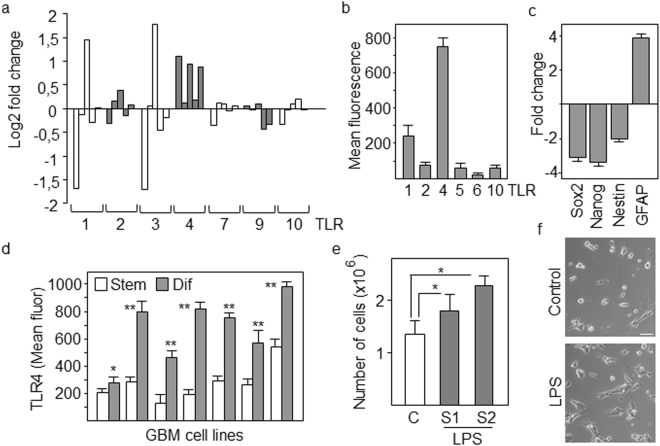
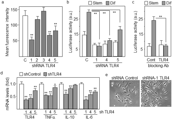
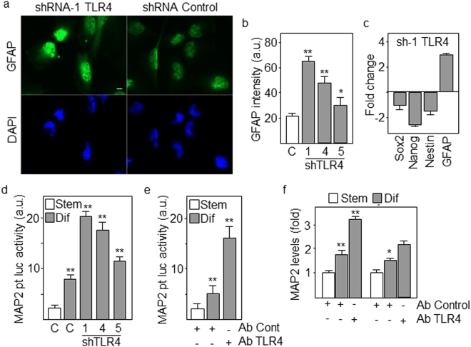
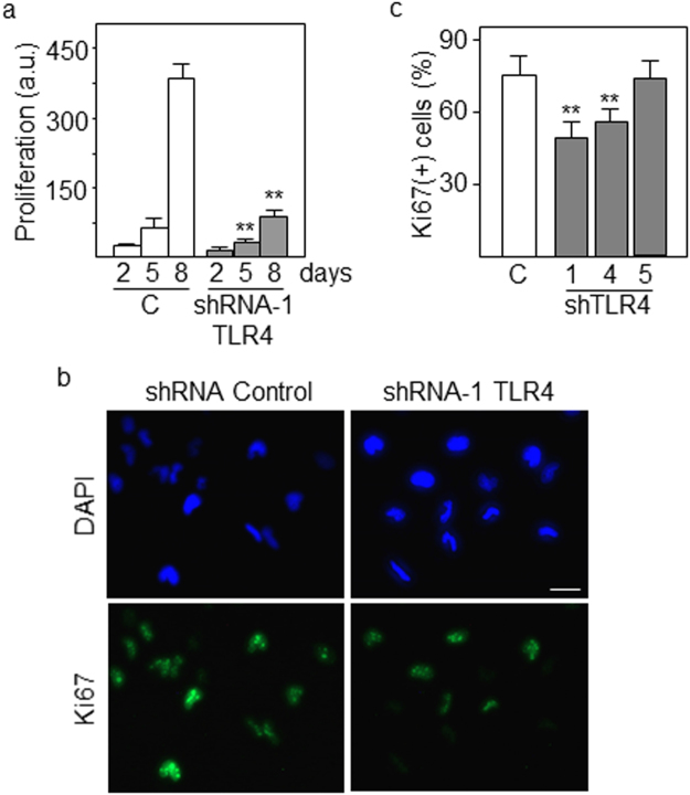
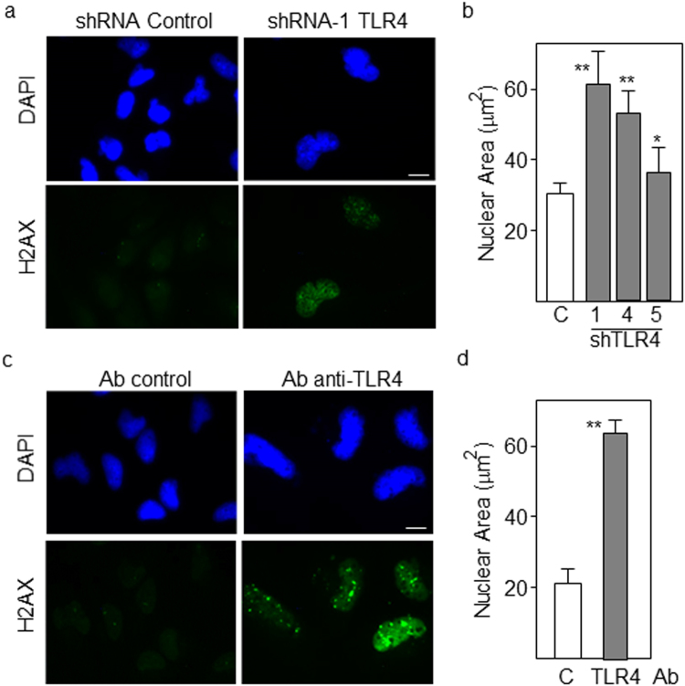
We have previously described that the NFκB pathway is upregulated during differentiation of glioblastoma stem-like cells (GSCs) which keeps differentiating GSCs in a proliferative astrocytic precursor state. However, extracellular signals and cellular mediators of this pathway are not clear yet. Here, we show that TLR4 is a key factor to promote NFκB activation in differentiating GSCs. TLR4 is upregulated during differentiation of GSCs and promotes transcriptional activation of NFκB as determined by luciferase-reporter assays and expression of NFκB target genes. Downregulation of TLR4 by shRNAs or blockade with anti-TLR4 specific antibodies drastically inhibited NFκB activity which promoted further differentiation and reduced proliferation of GSCs. We found that hyaluronic acid (HA), a main component of brain extracellular matrix, triggers the TLR4-NFκB pathway in differentiating GSCs. Moreover, HA is synthesized and released by GSCs undergoing differentiation and leads to transcriptional activation of NFκB, which is inhibited following downregulation of TLR4 or blockade of HA synthesis. Thus, we have demonstrated that during the process of differentiation, GSCs upregulate TLR4 and release the TLR4 ligand HA, which activates the TLR4-NFκB signaling pathway. This strategy may efficiently be used by differentiating GSCs to maintain their proliferative potential and consequently their tumorigenic capacity.


Conflict of interest statement

The authors declare no competing interests.

Figures

Figure 1
Figure 1
TLR4 is upregulated in differentiating GSCs. (a) Fold changes in the levels of TLR genes in differentiating compared with stem-like cells as assessed by gene expression microarray. (b) Flow cytometry analyses of TLRs. The expression in differentiating cells relative to stem-like cells is represented. (c) GSCs were induced to differentiate in the presence of serum for 4 days. The expression of progenitor (Nestin, Sox2, Nanog) and lineage (GFAP) markers was analyzed by qPCR. The expression levels were represented as fold changes of differentiating cells compared with GSCs. All markers showed significant differences (p < 0.01). (d) Expression levels of TLR4 protein in different GBM cell lines as determined by flow cytometry. Asterisks represent significant differences (*p < 0.05, **p < 0.01) compared to stem-like cells. (e) Proliferation and (f) Morphology of GSCs after 4 days of differentiation in the presence of LPS from E. coli strains O111:B4 (S1) and O55:B5 (S2). *p < 0.05. Histograms represent the mean ± SD of three independent experiments. Scale bar: 50 μm.
Figure 2
Figure 2
Downregulation of TLR4 blocks differentiation-induced activation of NFκB. (a) Protein levels of TLR4 are significantly reduced (**p < 0.01) in GBM cells with three out of five non-overlapping TLR4-specific shRNAs as determined by flow cytometry. (b,c) NFκB transcriptional activity following differentiation of GSCs transfected with TLR4-specific shRNAs (b) or cultured in the presence of TLR4 blocking antibodies (c). (d) Downregulation of TLR4 levels produced a decrease in the expression of NFκB target cytokines as determined by real-time PCR. (e) Morphological changes in GSCs with reduced levels of TLR4 after 4 days of differentiation. Scale bar: 50 μm. Histograms represent the mean ± SD of three independent experiments. **p < 0.01. a.u., arbitrary units.
Figure 3
Figure 3
Downregulation of TLR4 accelerates differentiation of GSCs. (a,b) GSCs were transfected with the indicated TLR4-specific shRNAs and expression of GFAP after 8 days of differentiation was assessed by immunofluorescence. Fluorescence intensity was measured using ImageJ software. Scale bar: 20 μm. (c) Cells were transfected with control or TLR4-specific shRNAs and differentiated for 8 days. The expression of progenitor (Nestin, Sox2, Nanog) and lineage (GFAP) markers was analyzed by qPCR. The expression levels were represented as fold changes of cells transfected with TLR4 shRNA compared with control cells. All markers showed significant differences (p < 0.01). (d) Transcriptional activity of the MAP2 promoter in stem and differentiating cells following transfection with three TLR4-specific shRNAs. (e) Transcriptional activity of the MAP2 promoter in cells treated with blocking anti-TLR4 antibodies. (f) Expression levels of MAP2 mRNA in stem and differentiating cells treated with blocking antibody. Histograms represent the mean ± SD of three independent experiments. Asterisks in grey bars represent significant differences (*p < 0.05, **p < 0.01) compared to empty (control) bars. a.u., arbitrary units.
Figure 4
Figure 4
Proliferation of differentiating GSCs decreases following downregulation of TLR4. (a) GSCs were transfected with TLR4-specific shRNAs and proliferation was assessed at different time points of differentiation by using Alamar Blue reagent. a.u., arbitrary units. (b) Expression of the proliferation marker Ki67 and (c) quantification of Ki67-positive cells in GSCs with low levels of TLR4 following 5 days of differentiation. Cells were counterstained with DAPI. Histograms represent the mean ± SD of three independent experiments. Asterisks in grey bars represent significant differences (**p < 0.01) compared to empty (control) bars. Scale bar: 10 μm.
Figure 5
Figure 5
Differentiating GSCs acquire a senescent phenotype after downregulation of TLR4. (a) Detection of histone H2AX phosphorylation by immunofluorescence staining of GSCs transfected with TLR4-specific shRNAs or (c) treated with blocking anti-TLR4 antibodies after 5 days of differentiation. Histograms shown in (b) and (d) represent the nuclear area of DAPI-stained cells treated as described in (a) and (b) respectively. At least 20 nuclei were counted. Asterisks in grey bars represent significant differences (*p < 0.05, **p < 0.01) compared to empty (control) bars. Scale bar: 5 μm.
Figure 6
Figure 6
Hyaluronic acid triggers TLR4 signaling in differentiating GSCs. (a) Cells were cotransfected with the NFκB-luciferase reporter vector and TLR4-specific or control shRNAs in the presence or in the absence of hyaluronic acid (HA). Luciferase activity was determined after 48 h of incubation under stem or differentiation conditions. (b) Proliferation of cells in the presence of HA following downregulation of TLR4 as determined by Alamar blue. (c) HA levels present in the culture medium of stem and differentiating cells treated with the HA synthesis inhibitor, 4-MU by ELISA. (d) Proliferation of stem and differentiating cells following treatment with 4-MU. (e) Luciferase activity of cells transfected with the NFκB-reporter vector and treated with 4-MU. (f) Expression levels of IL-6 mRNA in differentiating GSCs treated with 4-MU by quantitative RT-PCR. Histograms represent the mean ± SD of three independent experiments. Asterisks represent significant differences (**p < 0.01). a.u., arbitrary units.

References

    1. Eyler CE, Rich JN. Survival of the fittest: cancer stem cells in therapeutic resistance and angiogenesis. J Clin Oncol. 2008;26:2839–2845. doi: 10.1200/JCO.2007.15.1829. - DOI - PMC - PubMed
    1. Shay JW, Roninson IB. Hallmarks of senescence in carcinogenesis and cancer therapy. Oncogene. 2004;23:2919–2933. doi: 10.1038/sj.onc.1207518. - DOI - PubMed
    1. Kawamata H, Tachibana M, Fujimori T, Imai Y. Differentiation-inducing therapy for solid tumors. Curr Pharm Des. 2006;12:379–385. doi: 10.2174/138161206775201947. - DOI - PubMed
    1. Campos B, et al. Differentiation therapy exerts antitumor effects on stem-like glioma cells. Clin Cancer Res. 2010;16:2715–2728. doi: 10.1158/1078-0432.CCR-09-1800. - DOI - PubMed
    1. Naugler WE, Karin M. NF-kappaB and cancer-identifying targets and mechanisms. Curr Opin Genet Dev. 2008;18:19–26. doi: 10.1016/j.gde.2008.01.020. - DOI - PMC - PubMed

Publication types

MeSH terms

LinkOut - more resources