

EWSR1-PATZ1 gene fusion may define a new glioneuronal tumor entity
- PMID: 29679497
- PMCID: PMC8028282
- DOI: 10.1111/bpa.12619
EWSR1-PATZ1 gene fusion may define a new glioneuronal tumor entity
Abstract
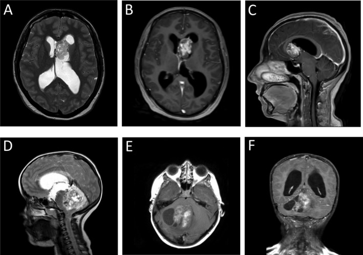
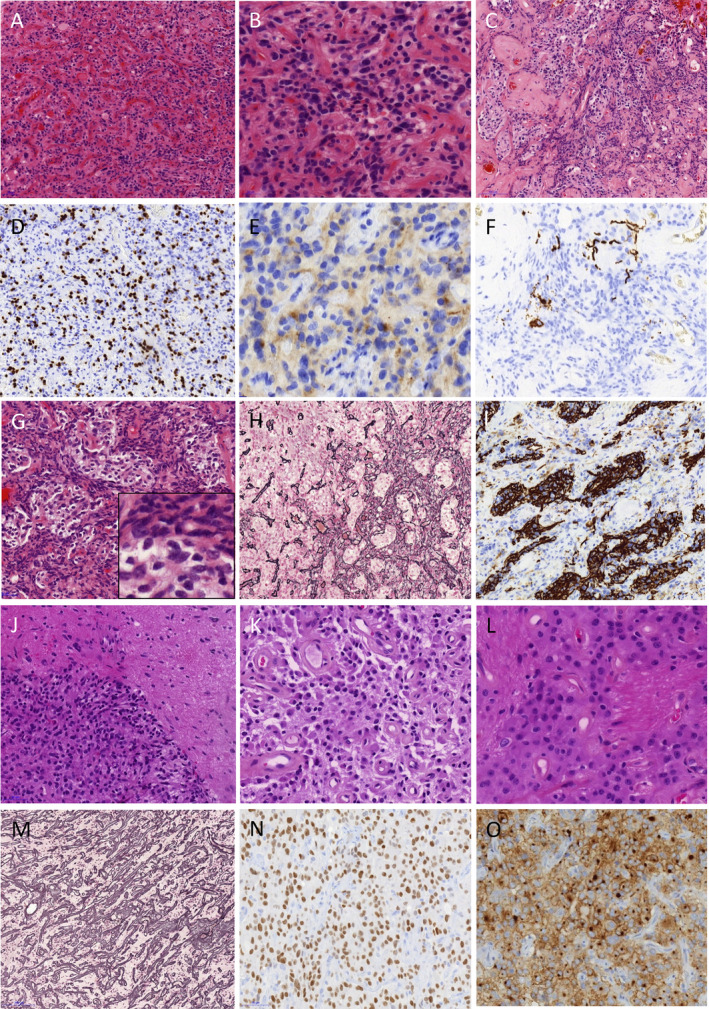
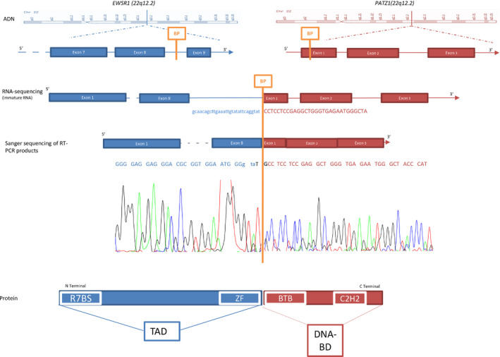
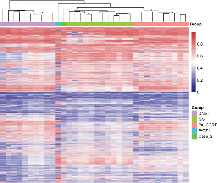
We investigated the challenging diagnostic case of a ventricular cystic glioneuronal tumor with papillary features, by RNA sequencing using the Illumina TruSight RNA Fusion panel. We did not retrieve the SLC44A1-PRKCA fusion gene specific for papillary glioneuronal tumor, but an EWSR1-PATZ1 fusion transcript. RT-PCR followed by Sanger sequencing confirmed the EWSR1-PATZ1 fusion. It matched with canonic EWSR1 fusion oncogene, juxtaposing the entire N-terminal transcriptional activation domain of EWSR1 gene and the C-terminal DNA binding domain of a transcription factor gene, PATZ1. PATZ1 protein belongs to the BTB-ZF (broad-complex, tramtrack and bric-à-brac -zinc finger) family. It directly regulates Pou5f1 and Nanog and is essential to maintaining stemness by inhibiting neural differentiation. EWSR1-PATZ1 fusion is a rare event in tumors: it was only reported in six round cell sarcomas and in three gliomas of three exclusively molecular studies. The first reported glioma was a BRAFV600E negative ganglioglioma, the second a BRAFV600E negative glioneuronal tumor, not otherwise specified and the third, very recently reported, a high grade glioma, not otherwise specified. In our study, forty BRAFV600E negative gangliogliomas were screened by FISH using EWSR1 break-apart probes. We performed methylation profiling for the index case and for seven out of the ten FISH positive cases. The index case clustered apart from other pediatric low grade glioneuronal entities, and specifically from the well-defined ganglioglioma methylation group. An additional pediatric intraventricular ganglioglioma clustered slightly more closely with ganglioglioma, but showed differences from the main ganglioglioma group and similarities with the index case. Both cases harbored copy number variations at the PATZ1 locus. EWSR1-PATZ1 gene fusion might define a new type of glioneuronal tumors, distinct from gangliogliomas.
Keywords: DNA methylation profiling; EWSR1; PATZ1; ganglioglioma; glioneuronal tumors.
© 2018 International Society of Neuropathology.
Figures
Similar articles
-
Glioneuronal tumors PATZ1-fused: clinico-molecular and DNA methylation signatures for a variety of morphological and radiological profiles.Acta Neuropathol Commun. 2025 May 24;13(1):114. doi: 10.1186/s40478-025-02037-5. Acta Neuropathol Commun. 2025. PMID: 40413488 Free PMC article.
-
PATZ1 fusions define a novel molecularly distinct neuroepithelial tumor entity with a broad histological spectrum.Acta Neuropathol. 2021 Nov;142(5):841-857. doi: 10.1007/s00401-021-02354-8. Epub 2021 Aug 21. Acta Neuropathol. 2021. PMID: 34417833 Free PMC article.
-
A novel LARGE1-AFF2 fusion expanding the molecular alterations associated with the methylation class of neuroepithelial tumors with PATZ1 fusions.Acta Neuropathol Commun. 2022 Feb 3;10(1):15. doi: 10.1186/s40478-022-01317-8. Acta Neuropathol Commun. 2022. PMID: 35115049 Free PMC article.
-
Neuroepithelial tumor with EWSR1::PATZ1 fusion: A literature review.J Neuropathol Exp Neurol. 2023 Oct 20;82(11):934-947. doi: 10.1093/jnen/nlad076. J Neuropathol Exp Neurol. 2023. PMID: 37804108 Review.
-
Round Cell Sarcoma with EWSR1-PATZ1 Gene Fusion in the Neck: Case Report and Review of the Literature.Laryngoscope. 2020 Dec;130(12):E833-E836. doi: 10.1002/lary.28554. Epub 2020 Mar 5. Laryngoscope. 2020. PMID: 32134119 Review.
Cited by
-
The spectrum of rare central nervous system (CNS) tumors with EWSR1-non-ETS fusions: experience from three pediatric institutions with review of the literature.Brain Pathol. 2021 Jan;31(1):70-83. doi: 10.1111/bpa.12900. Epub 2020 Nov 6. Brain Pathol. 2021. PMID: 32997853 Free PMC article. Review.
-
Giant-Cell Ependymoma of the Cervical Spinal Cord With Syringomyelia and Pathological Presentation: A Case Report and Review of the Literature.Cureus. 2022 Dec 31;14(12):e33174. doi: 10.7759/cureus.33174. eCollection 2022 Dec. Cureus. 2022. PMID: 36726917 Free PMC article.
-
Molecular profiling and targeted therapy in pediatric gliomas: review and consensus recommendations.Neuro Oncol. 2019 Aug 5;21(8):968-980. doi: 10.1093/neuonc/noz022. Neuro Oncol. 2019. PMID: 30805642 Free PMC article.
-
A Summary of the Inaugural WHO Classification of Pediatric Tumors: Transitioning from the Optical into the Molecular Era.Cancer Discov. 2022 Feb;12(2):331-355. doi: 10.1158/2159-8290.CD-21-1094. Epub 2021 Dec 17. Cancer Discov. 2022. PMID: 34921008 Free PMC article. Review.
-
Small round cell sarcomas.Nat Rev Dis Primers. 2022 Oct 6;8(1):66. doi: 10.1038/s41572-022-00393-3. Nat Rev Dis Primers. 2022. PMID: 36202860 Review.
References
-
- Chen X, Schulz‐Trieglaff O, Shaw R, Barnes B, Schlesinger F, Källberg M et al (2016) Manta: rapid detection of structural variants and indels for germline and cancer sequencing applications. Bioinformatics 32:1220–1222. - PubMed
Publication types
MeSH terms
Substances
LinkOut - more resources
Full Text Sources
Other Literature Sources
Medical
Research Materials
Miscellaneous
