Review
. 2018 Jul 20:664:152-167.
doi: 10.1016/j.gene.2018.04.048. Epub 2018 Apr 19.

MYH9: Structure, functions and role of non-muscle myosin IIA in human disease

Affiliations
Review

MYH9: Structure, functions and role of non-muscle myosin IIA in human disease

Alessandro Pecci et al. Gene. .

Abstract

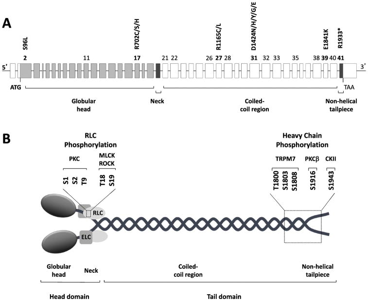
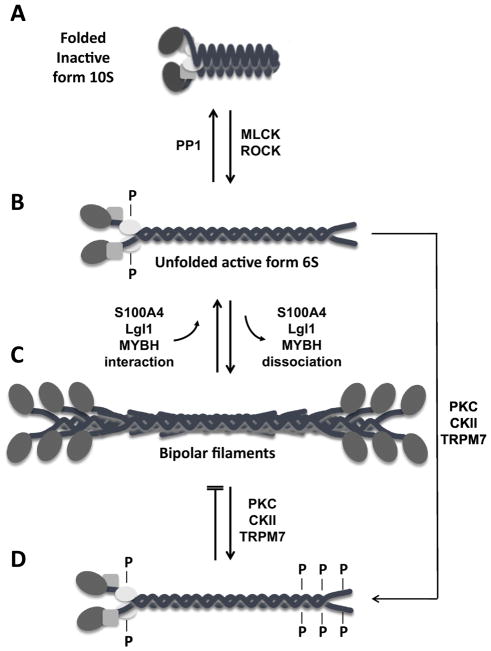
The MYH9 gene encodes the heavy chain of non-muscle myosin IIA, a widely expressed cytoplasmic myosin that participates in a variety of processes requiring the generation of intracellular chemomechanical force and translocation of the actin cytoskeleton. Non-muscle myosin IIA functions are regulated by phosphorylation of its 20 kDa light chain, of the heavy chain, and by interactions with other proteins. Variants of MYH9 cause an autosomal-dominant disorder, termed MYH9-related disease, and may be involved in other conditions, such as chronic kidney disease, non-syndromic deafness, and cancer. This review discusses the structure of the MYH9 gene and its protein, as well as the regulation and physiologic functions of non-muscle myosin IIA with particular reference to embryonic development. Moreover, the review focuses on current knowledge about the role of MYH9 variants in human disease.

Keywords: Actin-myosin cytoskeleton; Cell-cell adhesion; Class II myosin; Deafness; Inherited thrombocytopenia; Kidney disease; MYH9 gene; MYH9-related disease; Mouse models; Non-muscle myosin; Tumor suppressor.


Conflict of interest statement

CONFLICTS OF INTEREST

The authors declare no conflicts of interest.

Figures

Figure 1
Figure 1. MYH9 gene and protein organization
(A): Genomic structure of the MYH9 gene. MYH9 spans more than 106 kbp on chromosome 22q12.3 and is composed of 41 exons. The open reading frame spans exon 2 to exon 41 and encodes the non-muscle myosin heavy chain IIA, a protein of 1,960 amino acids. The numbers of the exons affected by the almost 90 different mutations responsible for MYH9-related disease (listed in Table 2) are indicated. Exons where hot spots of mutations are located, as well as the most frequent mutations, which account for approximately 70% of the MYH9-related disease families, are indicated in bold. (B): Schematic representation of non-muscle myosin IIA (NM IIA). NM IIA is a hexameric molecule consisting of a dimer of heavy chains, two regulatory light chains (RLC) and two essential light chains (ELC). Each heavy chain comprises the N-terminal head domain, which includes the motor (globular head, encoded by exons 1–19 of MYH9) and the neck (exon 20), and the C-terminal tail domain. The tail domain includes the long coiled-coil region (exons 21–40) and the short non-helical tailpiece (exon 41). Serine and threonine residues of RLC and heavy chains involved in phosphorylation, as well as the specific kinases, are indicated. PKC, protein kinase C; MLCK, myosin light chain kinase; ROCK, Rho-associated protein kinase; TRPM7, transient receptor potential melastatin 7; PKCβ, protein kinase Cβ; CKII, casein kinase II.
Figure 2
Figure 2. Regulation of non-muscle myosin IIA filament assembly
(A): Diagram of the folded, inactive 10S form of non-muscle myosin IIA (NM IIA) which is unable to assembly into filaments. Myosin light chain kinase (MLCK) and Rho-associated protein kinase (ROCK) phosphorylate RLC on serine 19 and threonine 18, allowing NM IIA to assume an unfolded active 6S conformation (B) and to assemble into bipolar filaments (C). The only known phosphatase that removes the phosphate group from these residues is protein phosphatase 1 (PP1). Assembly and disassembly of the bipolar filaments is also regulated by the interaction of NM IIA with S100A4, Lethal giant larvae (Lgl1), or myosin binding protein H (MYBH). (D) Phosphorylation of residues at the C-terminus of NM IIA by different kinases, such as protein kinase C (PKC), casein kinase 2 (CKII), and transient receptor potential melastatin 7 (TRPM7), either disassemblies the bipolar filaments or prevents their formation.
Figure 3
Figure 3. Expression of NM IIA in Developing Mouse Embryos
Sections of paraformaldehyde-fixed mouse embryos were stained with antibodies to NMHC IIA (green). NM IIA is widely distributed throughout the mouse embryos at E6.5 (A) and E11.5 (B). In E6.5 embryos, NM IIA is detected in all cells with similar staining intensity in both embryonic and extra-embryonic tissues (A). In E11.5 mouse embryos, different tissues and cells show marked variation in their staining intensity (B). In E16.5 mouse embryos, NM II-A is enriched in vasculature (endothelial cells) in brain (C), non-myocytes in heart (D), epithelial as well as interstitial mesenchymal cells in lung (E), and epithelial cells in intestine (F).
Figure 4
Figure 4. Abnormalities detectable at the examination of peripheral blood smears in patients with MYH9-related disease (MYH9-RD)
(A–C): Conventional panoptical May-Grünwald-Giemsa staining. (D–F): Immunofluorescence staining for the MYH9 protein (NMHC IIA). (A): Platelets of MYH9-RD patients are characterized by an extreme degree of macrocytosis: some platelets are even larger than erythrocytes (giant platelets). In (B) platelets of a healthy subjects are shown for comparison. (C): Aggregates of the MYH9 protein in the cytoplasm of neutrophil granulocytes may be identified after conventional staining of blood smears as faint basophilic (sky-blue) inclusion bodies, called “Döhle-like” bodies (arrow). (D–E): Immunofluorescence staining with antibodies to NMHC IIA allows to clearly detect the typical NMHC IIA aggregates in the cytoplasm of granulocytes of MYH9-RD patients and a definite diagnosis of the disorder. In (F) the distribution of the MYH9 protein in a granulocyte of a healthy individual is shown for comparison. The MYH9 genotypes of each individual are indicated in each image. WT, wild type. Scale bars correspond to 10 μm.

References

    1. Adelstein RS, Conti MA. Phosphorylation of platelet myosin increases actin-activated myosin ATPase activity. Nature. 1975;256:597–598. - PubMed
    1. Badirou I, Pan J, Souquere S, Legrand C, Pierron G, Wang A, Eckly A, Roy A, Gachet C, Vainchenker W, Chang Y, Léon C. Distinct localizations and roles of non-muscle myosin II during proplatelet formation and platelet release. J Thromb Haemost. 2015;13:851–859. - PubMed
    1. Baird MA, Billington N, Wang A, Adelstein RS, Sellers JR, Fischer RS, Waterman CM. Local pulsatile contractions are an intrinsic property of the myosin 2A motor in the cortical cytoskeleton of adherent cells. Mol Biol Cell. 2017;28:240–251. - PMC - PubMed
    1. Balduini A, Pallotta I, Malara A, Lova P, Pecci A, Viarengo G, Balduini CL, Torti M. Adhesive receptors, extracellular proteins and myosin IIA orchestrate proplatelet formation by human megakaryocytes. J Thromb Haemost. 2008;6:1900–1907. - PubMed
    1. Balduini CL, Pecci A, Noris P. Diagnosis and management of inherited thrombocytopenias. Semin Thromb Hemost. 2013;39:161–171. - PubMed

MeSH terms

Supplementary concepts