Review
. 2018 Jul 17;115(2):190-202.
doi: 10.1016/j.bpj.2018.03.028. Epub 2018 Apr 18.

Rediscovering Bacteria through Single-Molecule Imaging in Living Cells

Affiliations
Review

Rediscovering Bacteria through Single-Molecule Imaging in Living Cells

Achillefs N Kapanidis et al. Biophys J. .

Abstract

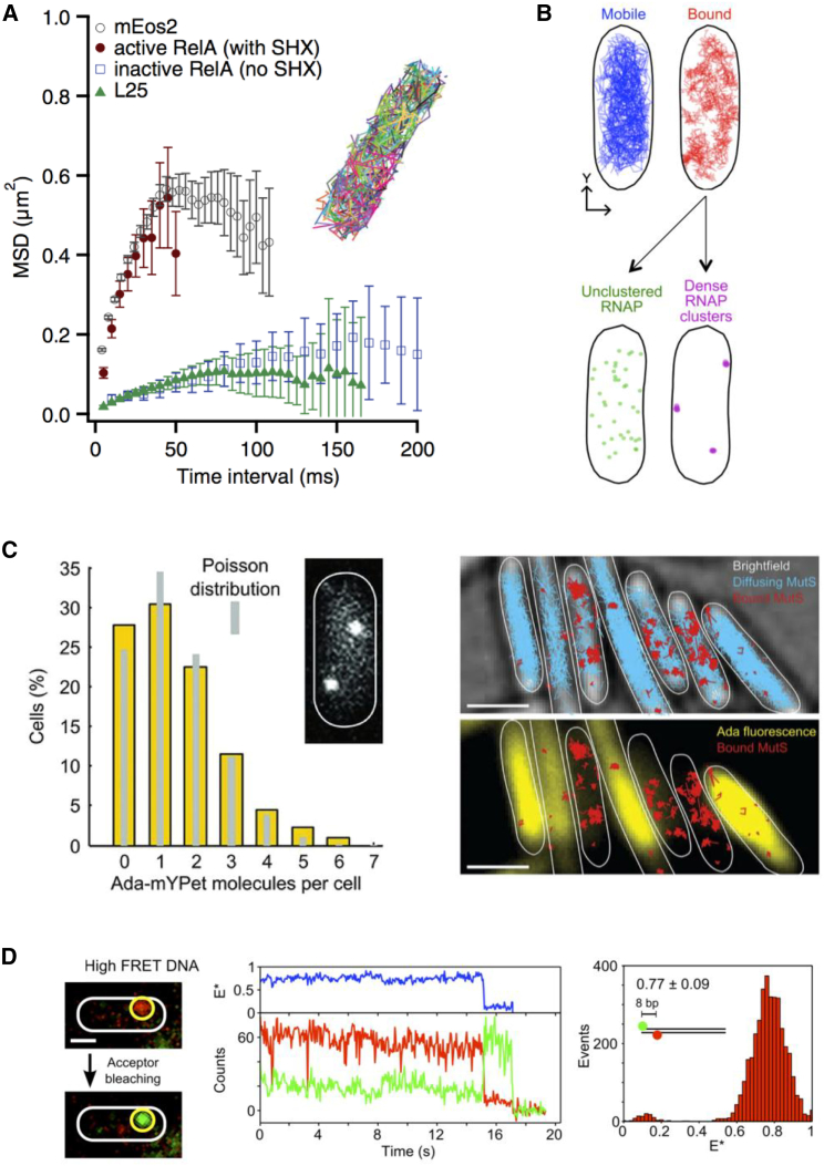
Bacteria are microorganisms central to health and disease, serving as important model systems for our understanding of molecular mechanisms and for developing new methodologies and vehicles for biotechnology. In the past few years, our understanding of bacterial cell functions has been enhanced substantially by powerful single-molecule imaging techniques. Using single fluorescent molecules as a means of breaking the optical microscopy limit, we can now reach resolutions of ∼20 nm inside single living cells, a spatial domain previously accessible only by electron microscopy. One can follow a single bacterial protein complex as it performs its functions and directly observe intricate cellular structures as they move and reorganize during the cell cycle. This toolbox enables the use of in vivo quantitative biology by counting molecules, characterizing their intracellular location and mobility, and identifying functionally distinct molecular distributions. Crucially, this can all be achieved while imaging large populations of cells, thus offering detailed views of the heterogeneity in bacterial communities. Here, we examine how this new scientific domain was born and discuss examples of applications to bacterial cellular mechanisms as well as emerging trends and applications.


Figures

Figure 1
Figure 1
The path to single-molecule detection of proteins inside living bacterial cells. A look at the evolution of imaging bacterial proteins using fluorescent protein fusions is shown. GFP was first developed as a biological probe for gene expression and was used on bacterial populations. Soon thereafter, fluorescence microscopy was focusing on single bacterial cells (10) as well as the subcellular distribution of proteins because there was adequate spatial resolution to do this. In 2006, it became possible to visualize single fluorescent protein fusions (using the Venus-YFP variant (23)) in cells with only a few copies of the protein of interest, and in 2008, the single-molecule detection capability was combined with photoactivation and tracking to study proteins of any copy number inside living bacterial cells (both nonactivated (P) and activated (FP) proteins are represented). To see this figure in color, go online.
Figure 2
Figure 2
Single-molecule fluorescence detection inside living bacteria. (A) A genetic construct occasionally produces a rare protein fusion that localizes on the inner bacterial membrane, which slows down its diffusion and allows detection as a diffraction-limited spot. (B) Differential interference contrast and fluorescence images of two bacterial cells show the presence of two fluorescence spots above the autofluorescence background; these spots correspond to single YFP molecules. (C) A time-series analysis of protein expression at the single-molecule level is shown. Each protein expression event persists for a significant time, likely because of the rate-limiting steps of fluorescence development in the YFP fluorophore. The figure is adapted from (23). To see this figure in color, go online.
Figure 3
Figure 3
Single-molecule localization and tracking. (A) Each single fluorescence molecule is detected as a 2D image with width similar or slightly larger than that of the point spread function (PSF) of the microscope (provided that relatively little motion of the fluorescent molecule occurs during the illumination time during the frame exposure). This image can be fitted with a Gaussian function, and its center can be identified with a precision that depends mainly on the number of photons per molecule per frame. (B) Molecules in the cell can be localized, and their motions can be tracked (lower panel). Because of the small dimensions of a bacterial cell relative to the size of the PSF (see example of an E. coli cell with just three molecules in top panel), the presence of many fluorescent molecules leads to a “crowded” situation that does not allow imaging of constantly fluorescent proteins (i.e., autofluorescing without photoactivation) with moderate-to-high copy numbers. (C) The principle of photoactivated single-molecule tracking in live bacteria is shown. Proteins are labeled with photoactivatable fusions, which are initially dark and can be turned on stochastically and at very low density using 405 nm light (or ultraviolet light); the activated molecule can be tracked using 561 nm light until it is bleached, and the cycle continues until all molecules are activated, tracked, and bleached, leading to a map of all tracks (lower left panel). To see this figure in color, go online.
Figure 4
Figure 4
Main single-molecule fluorescence observables inside living bacteria using fluorescent proteins. The observables apply for both autofluorescent and photoactivatable proteins, although the latter will provide higher statistics for counting and tracking. (A) The number of localizations per cell is shown, corresponding loosely to the protein copy number per cell. (B) Molecular mobility can be examined using plots of mean-square displacement (MSD) (pictured) or the cumulative distribution function (which plots the cumulative probability of finding a molecule within a certain distance after a certain time); this information can also be converted into apparent diffusion coefficients per track. In the example, a DNA polymerase (Pol1) in fixed cells shows no significant motion, whereas in live cells, it shows significant displacements until the confinement effects cause saturation of the MSDs; a smaller DNA-binding protein, Fis, shows faster motion. (C) Track location can be examined relative to the cell boundaries, relative to all other tracks (as pictured), relative to tracks with the same or different mobility, and relative to cell landmarks monitored in a different detection channel. (D) Time-series analysis of individual tracks can provide information about interaction (binding) kinetics identified by changes in the molecular mobility. In the example, a DNA polymerase molecule identifies its target, performs DNA synthesis, and resume its target search. Each step is 15 ms. Scale bars, 500 nm. The example figures are taken from (50). To see this figure in color, go online.
Figure 5
Figure 5
Applications of single-molecule imaging in living cells. (A) Using diffusion standards, it was shown that RelA diffusion, when inactive, is slow, matching that of ribosomes. The figure is adapted from (48). (B) The spatial profile of RNA polymerase shows that actively transcribed genes tends to be present in the nucleoid periphery. The figure is adapted from (51). (C) Single-molecule imaging of transcription factor Ada shows that some cells do not contain any molecule of this factor (left), delaying DNA-damage responses. This heterogeneity is also reflected in the diffusion profile of the MutS protein (right), which recognizes DNA mismatches that form when Ada is not present to repair damaged DNA molecules. There are many more DNA-bound MutS molecules (reflecting the presence of mismatches) in cells with low Ada content compared to cells in which Ada is abundant. The figure is adapted from (55). (D) Electroporated DNA can provide measurements of single-molecule FRET and distances within living cells. The figure is reproduced with permission from (69). To see this figure in color, go online.

References

    1. Selvin P.R., Ha T., editors. Single-Molecule Techniques: A Laboratory Manual. Cold Spring Harbor; New York: 2008.
    1. Xie X.S., Choi P.J., Lia G. Single-molecule approach to molecular biology in living bacterial cells. Annu. Rev. Biophys. 2008;37:417–444. - PubMed
    1. Hell S.W. Far-field optical nanoscopy. Science. 2007;316:1153–1158. - PubMed
    1. Stracy M., Uphoff S., Kapanidis A.N. In vivo single-molecule imaging of bacterial DNA replication, transcription, and repair. FEBS Lett. 2014;588:3585–3594. - PubMed
    1. Cattoni D.I., Fiche J.B., Nollmann M. Single-molecule super-resolution imaging in bacteria. Curr. Opin. Microbiol. 2012;15:758–763. - PubMed

Publication types