

Midtarsal locking, the windlass mechanism, and running strike pattern: A kinematic and kinetic assessment
- PMID: 29680311
- PMCID: PMC5944854
- DOI: 10.1016/j.jbiomech.2018.04.010
Midtarsal locking, the windlass mechanism, and running strike pattern: A kinematic and kinetic assessment
Abstract
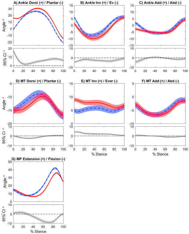
Changes in running strike pattern affect ankle and knee mechanics, but little is known about the influence of strike pattern on the joints distal to the ankle. The purpose of this study was to explore the effects of forefoot strike (FFS) and rearfoot strike (RFS) running patterns on foot kinematics and kinetics, from the perspectives of the midtarsal locking theory and the windlass mechanism. Per the midtarsal locking theory, we hypothesized that the ankle would be more inverted in early stance when using a FFS, resulting in decreased midtarsal joint excursions and increased dynamic stiffness. Associated with a more engaged windlass mechanism, we hypothesized that a FFS would elicit increased metatarsophalangeal joint excursions and negative work in late stance. Eighteen healthy female runners ran overground with both FFS and RFS patterns. Instrumented motion capture and a validated multi-segment foot model were used to analyze midtarsal and metatarsophalangeal joint kinematics and kinetics. During early stance in FFS the ankle was more inverted, with concurrently decreased midtarsal eversion (p < 0.001) and abduction excursions (p = 0.003) but increased dorsiflexion excursion (p = 0.005). Dynamic midtarsal stiffness did not differ (p = 0.761). During late stance in FFS, metatarsophalangeal extension was increased (p = 0.009), with concurrently increased negative work (p < 0.001). In addition, there was simultaneously increased midtarsal positive work (p < 0.001), suggesting enhanced power transfer in FFS. Clear evidence for the presence of midtarsal locking was not observed in either strike pattern during running. However, the windlass mechanism appeared to be engaged to a greater extent during FFS.
Keywords: Forefoot strike; Metatarsophalangeal joint; Midfoot; Multi-segment foot; Rearfoot strike.
Copyright © 2018 Elsevier Ltd. All rights reserved.
Conflict of interest statement
No conflicts of interest to report
Figures
 FFS,
FFS,
 RFS)
RFS) FFS,
RFS)
FFS,
RFS)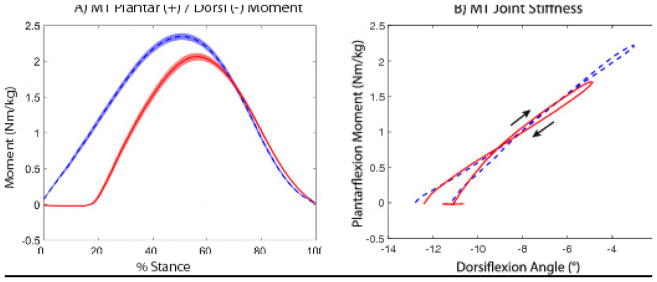 FFS,
RFS)
FFS,
RFS)References
-
- Alexander R, Ker R, Bennet M, Bibby S, Kester R. The spring in the arch of the human foot. Nature. 1987;325:147–149. - PubMed
-
- Andrade AG, Polese JC, Paolucci LA, Menzel H-JK, Teixeira-Salmela LF. Functional data analyses for the assessment of joint power profiles during gait of stroke subjects. Journal of applied biomechanics. 2014:30. - PubMed
-
- Ardigo L, Lafortuna C, Minetti A, Mognoni P, Saibene F. Metabolic and mechanical aspects of foot landing type, forefoot and rearfoot strike, in human running. Acta Physiologica Scandinavica. 1995;155:17–22. - PubMed
-
- Blackwood CB, Yuen TJ, Sangeorzan BJ, Ledoux WR. The midtarsal joint locking mechanism. Foot & ankle international. 2005;26:1074–1080. - PubMed
-
- Bruening DA, Cooney KM, Buczek FL. Analysis of a kinetic multi-segment foot model part II: kinetics and clinical implications. Gait & posture. 2012a;35:535–540. - PubMed
Publication types
MeSH terms
Grants and funding
LinkOut - more resources
Full Text Sources
Other Literature Sources
Miscellaneous
