Review
. 2018 Aug;59(3):253-268.
doi: 10.1007/s13353-018-0444-7. Epub 2018 Apr 21.

Splicing mutations in human genetic disorders: examples, detection, and confirmation

Affiliations
Review

Splicing mutations in human genetic disorders: examples, detection, and confirmation

Abramowicz Anna et al. J Appl Genet. 2018 Aug.

Erratum in

Abstract

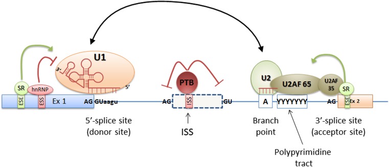
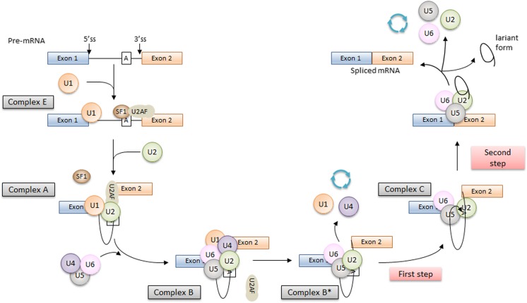
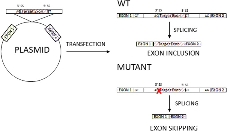
Precise pre-mRNA splicing, essential for appropriate protein translation, depends on the presence of consensus "cis" sequences that define exon-intron boundaries and regulatory sequences recognized by splicing machinery. Point mutations at these consensus sequences can cause improper exon and intron recognition and may result in the formation of an aberrant transcript of the mutated gene. The splicing mutation may occur in both introns and exons and disrupt existing splice sites or splicing regulatory sequences (intronic and exonic splicing silencers and enhancers), create new ones, or activate the cryptic ones. Usually such mutations result in errors during the splicing process and may lead to improper intron removal and thus cause alterations of the open reading frame. Recent research has underlined the abundance and importance of splicing mutations in the etiology of inherited diseases. The application of modern techniques allowed to identify synonymous and nonsynonymous variants as well as deep intronic mutations that affected pre-mRNA splicing. The bioinformatic algorithms can be applied as a tool to assess the possible effect of the identified changes. However, it should be underlined that the results of such tests are only predictive, and the exact effect of the specific mutation should be verified in functional studies. This article summarizes the current knowledge about the "splicing mutations" and methods that help to identify such changes in clinical diagnosis.

Keywords: Pre-mRNA splicing; Spliceosome; Splicing enhancers and silencers; Splicing mutation.


Conflict of interest statement

Conflict of interest

The authors declare that they have no conflict of interest.

Ethical approval

This article does not contain any studies with human participants or animals performed by any of the authors.

Figures

Fig. 1
Fig. 1
The schematic localization of the cis and trans splicing elements. The cis elements are the DNA sequences that include donor (5′) and acceptor (3′) splice sites, branch point and polypyrimidine tract sequences, and splicing silencers and enhancers. Donor and acceptor sites are evolutionary conserved and are usually defined by GT and AG nucleotides at the 5′ and 3′ ends of the intron, respectively. The branch site and the polypyrimidine tract sequences are highly degenerated and together with donor and acceptor sites are recognized by the elements of the splicing complex called spliceosome. Spliceosome proteins together with splicing repressors and activators recognize cis splicing elements and are called trans-acting elements
Fig. 2
Fig. 2
The scheme of the splicing process. The splicing process is performed in two steps. The first step is the recognition of the splicing sites at intron/exon junctions, and the second one is the intron removal and exon ends joining. During the splicing process, four complexes between the pre-mRNA and spliceosome are formed. The first one is the early complex (E). The U1 snRNP recognizes and binds to the complementary AG-GU sequence at the donor splice site (5′ end of the intron). In the same time, the SF1 protein binds to the branch point. The SF1 is recognized and bound by the U2AF65 protein that also binds to the polypyrimidine sequence located between the branch point and 3′ end of the intron. Then, the SF1 is displaced from the branch point by the U2 snRNP, and the ATP-dependent (A) complex is formed. The interaction between the branch point and the U2snRNP protein is stabilized by specific RNA helicases (Prp5 and Sub2), and this is a signal for the recruitment of U4/5/6 tri-snRNP and formation of the B complex (pre-catalytic spliceosome). Further action of additional RNA helicases leads to change of spliceosome conformation that leads to the release of U1 and U4 snRNPs, the interaction between U6 with U2 snRNP, and the formation of a pre-mRNA loop and the C complex. In this complex, two transesterification reactions take place, intron is removed, and ends of exon are joined (Fredericks et al. ; Tazi et al. 2009)
Fig. 3
Fig. 3
The types of splicing mutations. According to Wimmer et al. (2007), splicing mutations can be briefly divided into five categories: (1) splicing mutations within the canonical splice sites leading to whole exon skipping, (2) deep intronic variants creating new splice sites resulting in the inclusion of cryptic exons, (3) exonic single nucleotide variants creating new splice sites that result in the loss of an exon fragment, (4) variants in the canonical splice sites resulting in the usage of the cryptic exonic or intronic splice site that leads to the inclusion of an intron fragment or exon fragment skipping, and (5) mutations within the exon, usually leading to ESE disruption, resulting in the entire exon skipping
Fig. 4
Fig. 4
The principles of minigene assay. In the minigene assay, the amplified fragment of the analyzed gene, e.g., specific exon with surrounding intronic sequences with and without mutations, is cloned into a special expression plasmid enabling the analysis the pre-mRNA splicing (see description in text)

References

    1. Aoyama Y, Sasai H, Abdelkreem E, Otsuka H, Nakama M, Kumar S, Fukao T. A novel mutation (c.121-13T>A) in the polypyrimidine tract of the splice acceptor site of intron 2 causes exon 3 skipping in mitochondrial acetoacetyl-CoA thiolase gene. Mol Med Rep. 2017;15:3879–3884. doi: 10.3892/mmr.2017.6434. - DOI - PubMed
    1. Axelrod FB, Liebes L, Gold-von Simson G, et al. Kinetin improves IKBKAP mRNA splicing in patients with familial dysautonomia. Pediatr Res. 2011;70(5):480–483. doi: 10.1203/PDR.0b013e31822e1825. - DOI - PMC - PubMed
    1. Baralle D, Baralle M. Splicing in action: assessing disease causing sequence changes. J Med Genet. 2005;42(10):737–748. doi: 10.1136/jmg.2004.029538. - DOI - PMC - PubMed
    1. Bergsma AJ, van der Wal E, Broeders M, van der Ploeg AT, Pim Pijnappel WWM. Alternative splicing in genetic diseases: improved diagnosis and novel treatment options. Int Rev Cell Mol Biol. 2018;335:85–141. doi: 10.1016/bs.ircmb.2017.07.008. - DOI - PubMed
    1. Brendel V, Xing L, Zhu W. Gene structure prediction from consensus spliced alignment of multiple ESTs matching the same genomic locus. Bioinformatics. 2004;20:1157–1169. doi: 10.1093/bioinformatics/bth058. - DOI - PubMed