Review
. 2018 Jun;5(2):R23-R36.
doi: 10.1530/ERP-18-0016. Epub 2018 Mar 16.

EDUCATIONAL SERIES IN CONGENITAL HEART DISEASE: Congenital left-sided heart obstruction

Affiliations
Review

EDUCATIONAL SERIES IN CONGENITAL HEART DISEASE: Congenital left-sided heart obstruction

Michelle Carr et al. Echo Res Pract. 2018 Jun.

Abstract

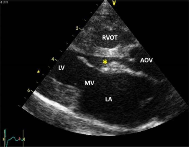
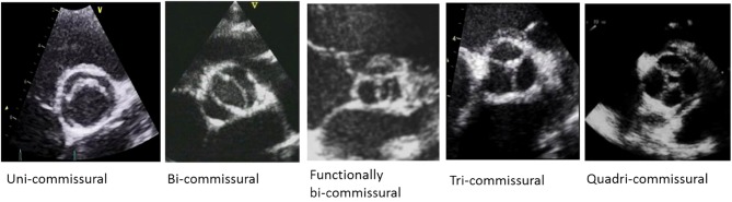
Congenital obstruction of the left ventricular outflow tract remains a significant problem and multilevel obstruction can often coexist. Obstruction can take several morphological forms and may involve the subvalvar, valvar or supravalvar portion of the aortic valve complex. Congenital valvar stenosis presenting in the neonatal period represents a spectrum of disorders ranging from the hypoplastic left heart syndrome to almost normal hearts. Treatment options vary dependent on the severity of the left ventricular outflow tract obstruction (LVOTO) and the variable degree of left ventricular hypoplasia as well as the associated lesions such as arch hypoplasia and coarctation.

Keywords: aortic stenosis; bicuspid aortic valve; coarctation of the aorta; congenital heart defects; left ventricular outflow tract obstruction.


Figures

Figure 1
Figure 1
Circular fibromuscular shelf seen in 2D parasternal long axis view. AOV, aortic valve; LA, left atrium; LV, left ventricle; MV, mitral valve; RVOT, right ventricular outflow tract.
Figure 2
Figure 2
Circular subaortic membrane (arrows) adherent to aortic valve (AOV) visualised in transoesophageal 2D imaging (A) and colour flow mapping (B) in long axis view (130°). LVOT, left ventricular outflow tract; RVOT, right ventricular outflow tract.
Figure 3
Figure 3
Fibromuscular semicircular shelf (arrows) in real-time 3D transthoracic echocardiography as would be seen by a surgeon (surgeon’s view). LVOT, left ventricular outflow tract; MV, mitral valve; TV, tricuspid valve.
Figure 4
Figure 4
LV outflow tract obstruction caused by hypertrophied subaortic conus in 2D parasternal long axis view. AOV, aortic valve; LA, left atrium; LV, left ventricle; MV, mitral valve; RVOT, right ventricular outflow tract.
Figure 5
Figure 5
LV outflow tract obstruction caused by posterior deviation of infundibular septum (asterisk) in association with ventricular septal defect (VSD, arrow), bicuspid aortic valve (AOV) in a newborn baby with interrupted aortic arch. 2D image in parasternal long axis view. LA, left atrium; LV, left ventricle; RVOT, right ventricular outflow tract.
Figure 6
Figure 6
LV outflow tract obstruction in patient with HOCM. Obstruction caused dynamic LV outflow tract obstruction and chordal attachment from anterior mitral leaflet (asterisk). 2D parasternal long axis view (A) and real-time 3D small-volume transoesophageal echocardiography (B). RVOT, right ventricular outflow tract; AOV, aortic valve; LA, left atrium; LV, left ventricle.
Figure 7
Figure 7
Real-time 3D transthoracic echocardiography (A) with LV outflow tract obstruction visualised from LV apex (A) and surgeon`s view (B) demonstrating obstructive tissue (arrows) protruding to LV outflow tract (LVOT) during systole when mitral valve (MV) is completely closed. AOV, aortic valve; LV, left ventricle; RV, right ventricle.
Figure 8
Figure 8
An adolescent with HOCM. 2D parasternal long axis view with colour flow mapping (A) demonstrates flow turbulence within narrow LV outflow tract (LVOT) simultaneously with moderate mitral regurgitant jet diverted very posteriorly. M-mode technique (B) taken from same long axis view confirms systolic anterior motion (SAM) of usually elongated mitral leaflet or supporting chord (arrows). CW Doppler (C) indicates late systolic peak (arrows) confirming dynamic LV outflow tract obstruction. Another CW Doppler (D) displayed simultaneously very high mid-systolic peak from mitral regurgitation (white arrows) and late systolic peak from LV outflow tract (yellow arrows). LA, left atrium.
Figure 9
Figure 9
Four chamber (A) and parasternal long axis view (B) in neonate with critical aortic stenosis (asterisk) and endocardial fibroelastosis (EFE) seen as hyperechogenic layer within hypertrophied left ventricle (LV). LA, left atrium; RA, right atrium; RV, right ventricle.
Figure 10
Figure 10
Panel of morphological variants of aortic valves visualised in 2D transthoracic echocardiography in parasternal short axis view.
Figure 11
Figure 11
Bicuspid (bi-commissural) aortic valve with vertical orientation of effective orifice imaged by transoesophageal real-time 3D echocardiography. Purely bicuspid valve with vertical orientation of effective orifice and absence of raphe or fused commissure seen during systole (A) and diastole (B). Another example of bicuspid aortic valve demonstrates horizontal orientation of effective orifice during systole (C) and thick raphe within Valsalva sinuses at 2o’clock (arrow) seen within pockets of sinuses during diastolic closure (D).
Figure 12
Figure 12
Echocardiographic measurement of aortic root (AO) in parasternal long axis view: annulus, Valsalva sinuses, sinotubular junction and ascending aorta dimensions measured in a patient with Marfan syndrome. Measurements performed during systole, aortic annulus measured at the level of aortic leaflet hinge points. AO, aorta; LA, left atrium; LVOT, left ventricular outflow tract.
Figure 13
Figure 13
2D and colour flow mapping taken from high parasternal transthoracic approach (A) confirms optimal blood flow alignment for correct measurement of flow on CW Doppler (B). AO, ascendant aorta; LV, left ventricle.
Figure 14
Figure 14
2D parasternal long axis (A) and colour flow mapping (B) in a patient with severe narrowing at the level of sinotubular junction (arrows). AO, aorta; AOV, aortic valve; LA, left atrium; LV, left ventricle.
Figure 15
Figure 15
2D parasternal long axis (A) and colour flow mapping (B) in a patient with Williams-Beuren syndrome. Diffuse hypoplasia with markedly thickened aortic wall with flow turbulence occurring just at the level of sinotubular junction (B). AO, aorta; AOV, aortic valve; LA, left atrium; LV, left ventricle.
Figure 16
Figure 16
Schematic drawing of proximal and distal aortic arch, aortic isthmus, arterial duct or ligamentum (asterisk) and descending aorta. LCAR, left carotid artery; LPA, left pulmonary artery; PA, pulmonary trunk; RCAR, right carotid artery; RPA, right pulmonary artery; RSA, right subclavian artery. Reproduced, with permission, from Marek J, Fenton M & Khambadkone S (2015) Aortic arch anomalies: coarctation of the aorta and interrupted aortic arch. In Echocardiography in Pediatric and Congenital Heart Disease: From Fetus to Adult, 2nd edn, ch 21. Eds WW Lai, LL Mertens, MS Cohen, T Geva. Copyright 2015 Wiley-Blackwell.
Figure 17
Figure 17
Neonatal coarctation (COA) with severe transverse aortic arch hypoplasia from suprasternal view. Elongated hypoplastic arch with maximum of narrowing between left carotid artery (LCAR), left vertebral artery (LVA) and left subclavian artery (LSA), which is predominantly supplied by arterial duct (asterisk). BCT, brachiocephalic trunk; DAO, descendent aorta. Reproduced, with permission, from Marek J, Fenton M & Khambadkone S (2015) Aortic arch anomalies: coarctation of the aorta and interrupted aortic arch. In Echocardiography in Pediatric and Congenital Heart Disease: From Fetus to Adult, 2nd edn, ch 21. Eds WW Lai, LL Mertens, MS Cohen, T Geva. Copyright 2015 Wiley-Blackwell.
Figure 18
Figure 18
Imaging of COA from the suprasternal notch view. Coarctation ridge (COA) located ~5 mm below the origin of left subclavian artery. Descendent thoracic aorta (DAO) below obstruction is dilated. AOA, aortic arch. Reproduced, with permission, from Marek J, Fenton M & Khambadkone S (2015) Aortic arch anomalies: coarctation of the aorta and interrupted aortic arch. In Echocardiography in Pediatric and Congenital Heart Disease: From Fetus to Adult, 2nd edn, ch 21. Eds WW Lai, LL Mertens, MS Cohen, T Geva. Copyright 2015 Wiley-Blackwell.
Figure 19
Figure 19
Pulsed Doppler flow pattern in the abdominal aorta from subcostal sagittal view. Systolic wave form amplitude is low; antegrade diastolic flow indicates the obstruction proximal to sample volume. Reproduced, with permission, from Marek J, Fenton M & Khambadkone S (2015) Aortic arch anomalies: coarctation of the aorta and interrupted aortic arch. In Echocardiography in Pediatric and Congenital Heart Disease: From Fetus to Adult, 2nd edn, ch 21. Eds WW Lai, LL Mertens, MS Cohen, T Geva. Copyright 2015 Wiley-Blackwell.
Figure 20
Figure 20
Continuous wave Doppler through the aortic isthmus of a patient with severe COA. Note the high-velocity systolic amplitude (4.5 m/s) with continuous antegrade flow throughout diastole. Reproduced, with permission, from Marek J, Fenton M & Khambadkone S (2015) Aortic arch anomalies: coarctation of the aorta and interrupted aortic arch. In Echocardiography in Pediatric and Congenital Heart Disease: From Fetus to Adult, 2nd edn, ch 21. Eds WW Lai, LL Mertens, MS Cohen, T Geva. Copyright 2015 Wiley-Blackwell.
Figure 21
Figure 21
(A) Recurrent coarctation of aorta (reCOA) in a patient after left subclavian flap aortoplasty (Waldhausen procedure). Narrowing with flow acceleration seen by colour Doppler flow mapping at the level of origin of left subclavian artery, which was used to extend tubular aortic narrowing. (B) Stent implantation at the level of residual narrowing in a patient with recurrent coarctation (arrows). AOA, aortic arch; LCAR, left carotid artery; RPA, right pulmonary artery. Reproduced, with permission, from Marek J, Fenton M & Khambadkone S (2015) Aortic arch anomalies: coarctation of the aorta and interrupted aortic arch. In Echocardiography in Pediatric and Congenital Heart Disease: From Fetus to Adult, 2nd edn, ch 21. Eds WW Lai, LL Mertens, MS Cohen, T Geva. Copyright 2015 Wiley-Blackwell.

References

    1. Katz NM, Buckley MJ, Liberthson RR. Discrete membranous subaortic stenosis. Report of 31 patients, review of the literature, and delineation of management. Circulation 1977. 56 1034–1038. (10.1161/01.CIR.56.6.1034) - DOI - PubMed
    1. Gewillig M, Daenen W, Dumoulin M, Van der Hauwaert L. Rheologic genesis of discrete subvalvular aortic stenosis: a Doppler echocardiographic study. Journal of the American College of Cardiology 1992. 19 818–824. (10.1016/0735-1097(92)90524-Q) - DOI - PubMed
    1. Choi JY, Sullivan ID. Fixed subaortic stenosis: anatomical spectrum and nature of progression. British Heart Journal 1991. 65 280–286. (10.1136/hrt.65.5.280) - DOI - PMC - PubMed
    1. Brauner R, Laks H, Drinkwater DC, Jr, Shvarts O, Eghbali K, Galindo A. Benefits of early surgical repair in fixed subaortic stenosis. Journal of the American College of Cardiology 1997. 30 1835–1842. (10.1016/S0735-1097(97)00410-5) - DOI - PubMed
    1. Wright GB, Keane JF, Nadas AS, Bernhard WF, Castaneda AR. Fixed subaortic stenosis in the young: medical and surgical course in 83 patients. American Journal of Cardiology 1983. 52 830–835. (10.1016/0002-9149(83)90423-X) - DOI - PubMed

LinkOut - more resources