Review
. 2018 Apr 5;15(2):529-535.
doi: 10.1016/j.jor.2018.03.039. eCollection 2018 Jun.

Pediatric proximal femur fractures

Affiliations
Review

Pediatric proximal femur fractures

Brian L Dial et al. J Orthop. .

Abstract

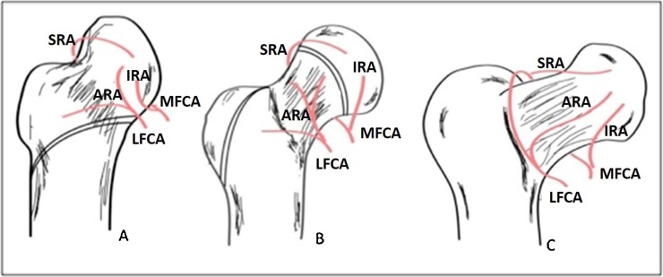
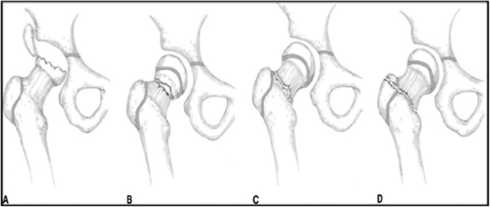
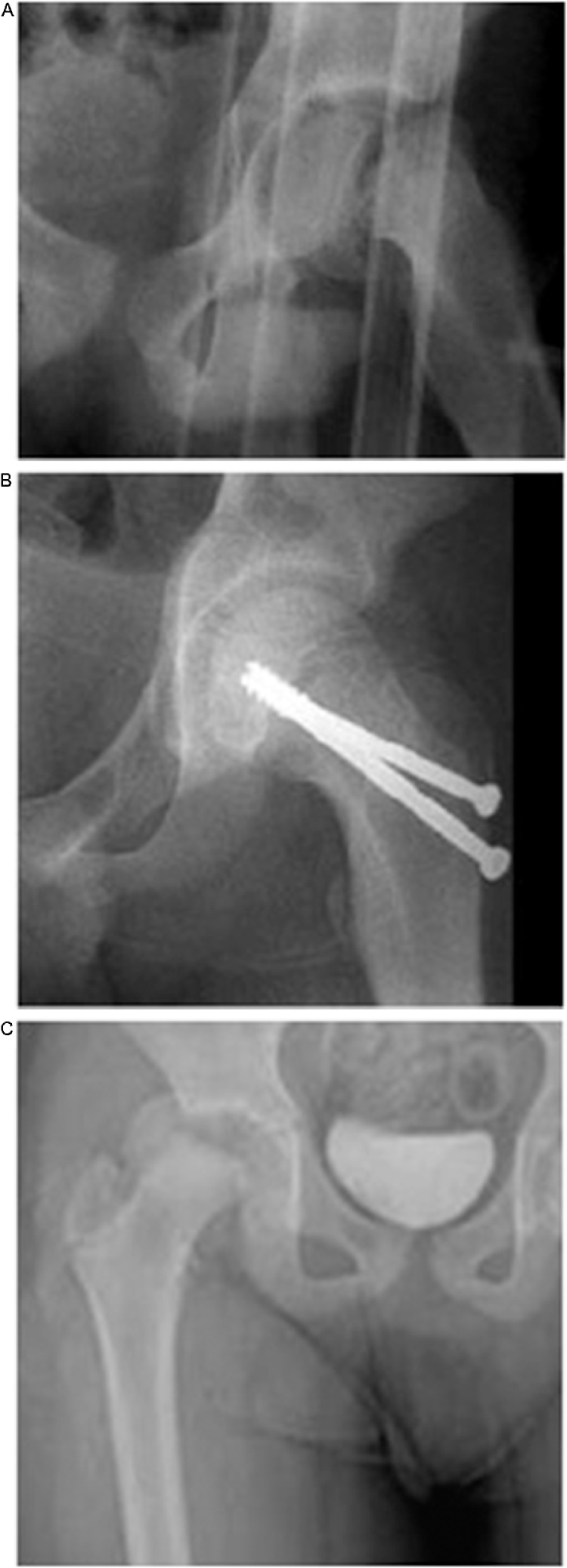
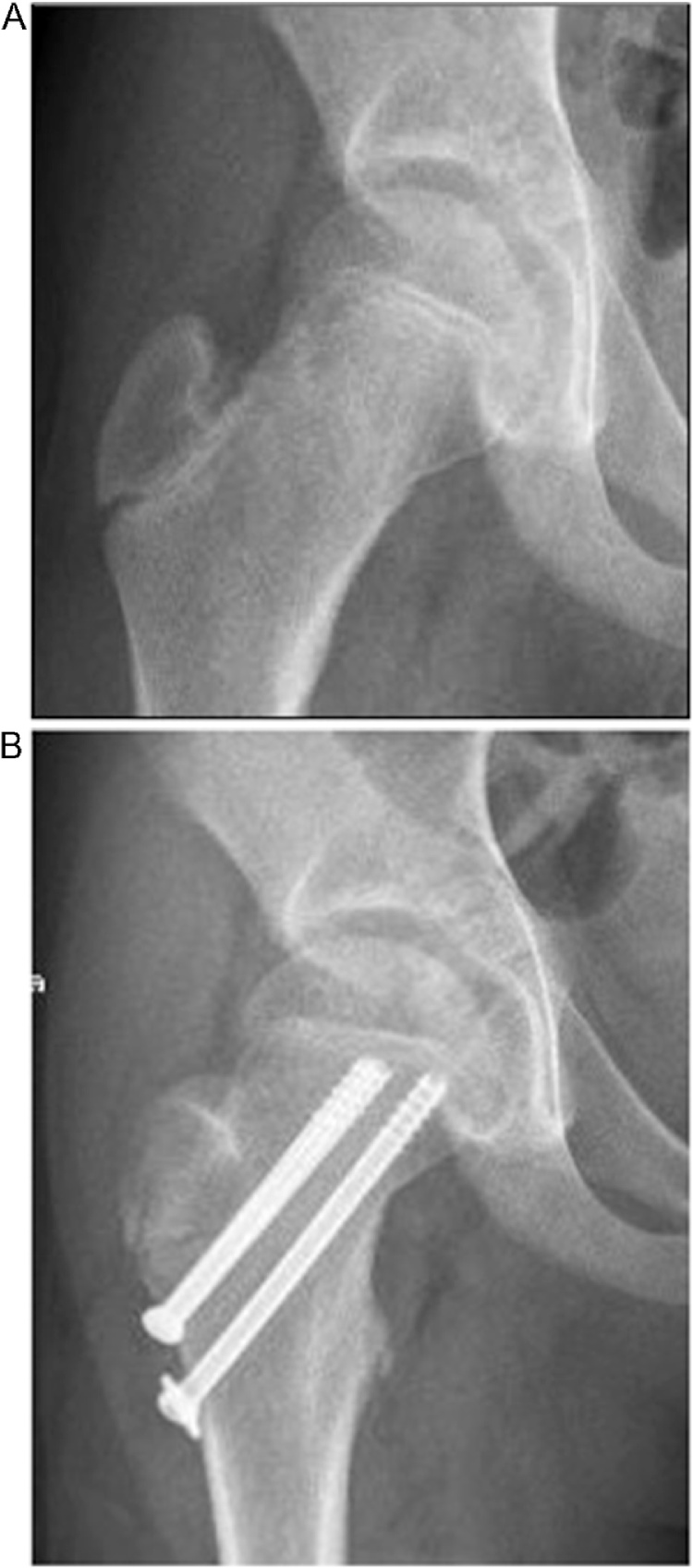
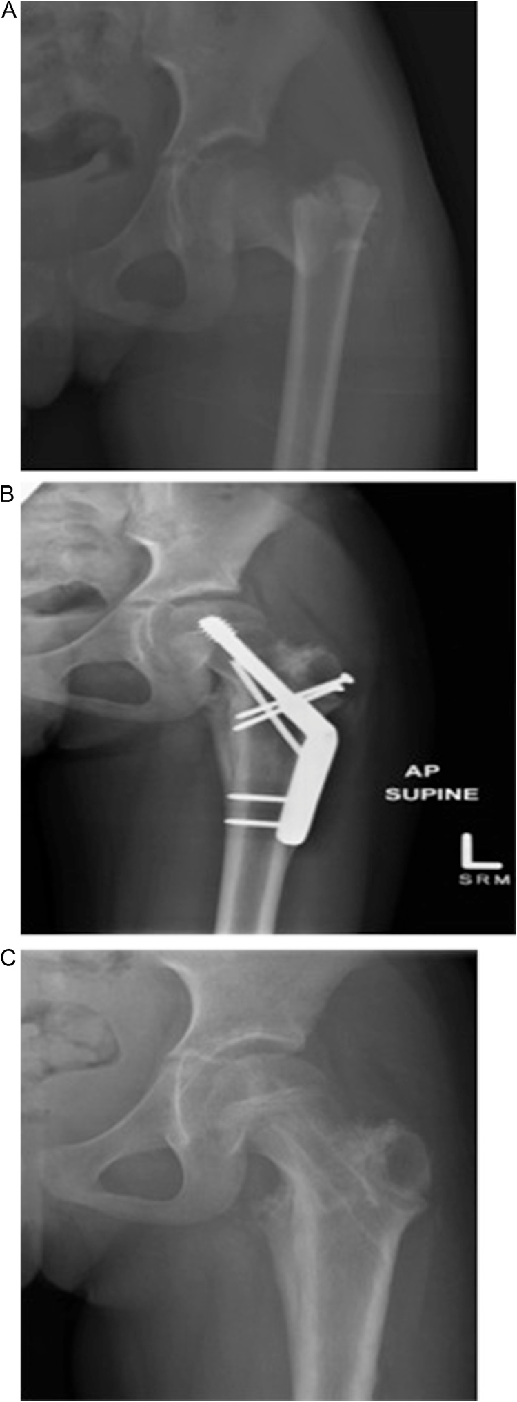
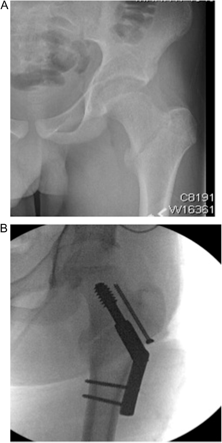
Pediatric proximal femur fractures are rare injuries resulting from high-energy trauma. The Delbet classification is used when describing these injuries, and associates fracture type to the development of avascular necrosis. Historically, casting was utilized in the treatment of these injuries, but high complication rates following this approach have changed the treatment modality to early and anatomic fixation. Complications associated with these injuries including avascular necrosis, non-union, coxa-vara, and premature physeal fusion. Achieving anatomic reduction and performing internal fixation within 24 h from time of injury has become the standard of care in the treatment of pediatric proximal femur fractures.

Keywords: Children hip fracture; Delbet classification; Pediatric femoral neck fracture; Pediatric hip fracture; Pediatric proximal femur fracture.


Figures

Fig. 1
Fig. 1
Vascular anatomy of the pediatric proximal femur. The image also depicts the progressive femoral neck-shaft angle from 150° in the newborn to 130° in the skeletally maturity. A, Stage 1 with tri-vessel supply. B, Stage 2, the ARA no longer supplies the femoral head due to the physis. C, Stage 3 the skeletally mature vascular supply. MFCA – medial femoral circumflex artery, LFCA – lateral femoral circumflex artery, SRA – superior retinacular artery, IRA – inferior retinacular artery, ARA – anterior retinacular artery.
Fig. 2
Fig. 2
The Delbet classification of pediatric proximal femur fractures. A, type 1 transepiphyseal fracture. B, type II transcervical fracture. C, type III basicervical fracture. D, type IV intertrochanteric fracture.
Fig. 3
Fig. 3
A) A 13 year-old boy who sustained a Delbet IA fracture during a snowmobiling accident. B) Two partially threaded screws were used to stabilize the fracture. These screws are inserted through the physis. C) For comparison, this is a radiograph of a child who experienced a Delbet IB fracture, the femoral head has dislocated from the acetabulum.
Fig. 4
Fig. 4
A) 12 year-old boy presents with a non-displaced transcervical fracture (Delbet II). B) Three 6.5 m m partially threaded screws were inserted in an inverted triangle configuration. The screws do not cross the physis, and a washer was used for further stabilization with the inferior screw.
Fig. 5
Fig. 5
A) Basicervical fracture (Delbet III) with an associated greater trochanter fracture in a 7 year-old girl following a MVC B) A pediatric sliding hip screw (SHS) was used for primary stabilization, and a second screw was used to control rotation. In addition, two screws were used to stabilize the greater trochanteric fracture. C) Following fracture healing the hardware was removed to prevent physeal arrest.
Fig. 6
Fig. 6
A) An intertrochanteric fracture (Delbet IV) in a patient approaching skeletal maturity. B) Intra-operative fluoroscopy showing a standard hip screw (SHS) was used for primary stabilization, and a partially threaded screw was inserted across the fracture site to prevent rotation.

References

    1. Ratliff A.H. Fractures of the neck of the femur in children. J Bone Jt Surg. 1962;44B(3) - PubMed
    1. Spence D., DiMauro J.P., Miller P.E., Glotzbecker M.P., Hedequist D.J., Shore B.J. Osteonecrosis after femoral neck fractures in children and adolescents: analysis of risk factors. J Pediatr Orthop. 2016;36(2):111–116. - PubMed
    1. Bali K., Sudesh P., Patel S., Kumar V., Saini U., Dhillon M.S. Pediatric femoral neck fractures: our 10 years of experience. Clin Orthop Surg. 2011;3(4):302–308. - PMC - PubMed
    1. Boardman M.J., Herman M.J., Buck B., Pizzutillo P. Hip fractures in children. JAAOS. 2009;17(3):162–173. - PubMed
    1. Trueta J. The normal vascular anatomy of the human femoral head during growth. J Bone Jt Surg Br. 1957;39-b(2):358–394. - PubMed

LinkOut - more resources