Clinical and Prognostic Value of PET/CT Imaging with Combination of 68Ga-DOTATATE and 18F-FDG in Gastroenteropancreatic Neuroendocrine Neoplasms
- PMID: 29681780
- PMCID: PMC5846381
- DOI: 10.1155/2018/2340389
Clinical and Prognostic Value of PET/CT Imaging with Combination of 68Ga-DOTATATE and 18F-FDG in Gastroenteropancreatic Neuroendocrine Neoplasms
Abstract
Background: To evaluate the clinical and prognostic value of PET/CT with combination of 68Ga-DOTATATE and 18F-FDG in gastroenteropancreatic neuroendocrine neoplasms (GEP-NENs).
Method: 83 patients of GEP-NENs who underwent 68Ga-DOTATATE and 18F-FDG PET/CT were enrolled between June 2013 and December 2016. Well-differentiated (WD) NETs are divided into group A (Ki-67 < 10%) and group B (Ki-67 ≥ 10%), and poorly differentiated (PD) NECs are defined as group C. The relationship between PET/CT results and clinicopathological characteristics was retrospectively investigated.
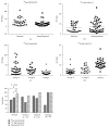
Result: For groups A/B/C, the sensitivities of 68Ga-DOTATATE and 18F-FDG were 78.8%/83.3%/37.5% and 52.0%/72.2%/100.0%. A negative correlation between Ki-67 and SUVmax of 68Ga-DOTATATE (R = -0.415; P ≤ 0.001) was observed, while a positive correlation was noted between Ki-67 and SUVmax of 18F-FDG (R = 0.683; P ≤ 0.001). 62.5% (5/8) of patients showed significantly more lesions in the bone if 68Ga-DOTATATE was used, and 22.7% (5/22) of patients showed more lymph node metastases if 18F-FDG was used.
Conclusions: The sensitivity of dual tracers was correlated with cell differentiation, and a correlation between Ki-67 and both SUVmax of PET-CTs could be observed. 68Ga-DOTATATE is suggested for WD-NET and 18F-FDG is probably suitable for patients with Ki-67 ≥ 10%.
Figures


Similar articles
-
[68Ga-DOTATATE and 18F-FDG PET/CT dual-modality imaging enhances precision of staging and treatment decision for gastroenteropancreatic neuroendocrine neoplasms].Nan Fang Yi Ke Da Xue Xue Bao. 2025 Jun 20;45(6):1212-1219. doi: 10.12122/j.issn.1673-4254.2025.06.10. Nan Fang Yi Ke Da Xue Xue Bao. 2025. PMID: 40579134 Free PMC article. Chinese.
-
Can complementary 68Ga-DOTATATE and 18F-FDG PET/CT establish the missing link between histopathology and therapeutic approach in gastroenteropancreatic neuroendocrine tumors?J Nucl Med. 2014 Nov;55(11):1811-7. doi: 10.2967/jnumed.114.142224. Epub 2014 Oct 14. J Nucl Med. 2014. PMID: 25315243
-
The Correlation Between [68Ga]DOTATATE PET/CT and Cell Proliferation in Patients With GEP-NENs.Mol Imaging Biol. 2019 Oct;21(5):984-990. doi: 10.1007/s11307-019-01328-3. Mol Imaging Biol. 2019. PMID: 30796708
-
Ga-68 DOTA-peptides and F-18 FDG PET/CT in patients with neuroendocrine tumor: A review.Clin Imaging. 2020 Nov;67:113-116. doi: 10.1016/j.clinimag.2020.05.035. Epub 2020 Jun 9. Clin Imaging. 2020. PMID: 32559681 Review.
-
Gastroenteropancreatic neuroendocrine tumours (GEP-NET) - Imaging and staging.Best Pract Res Clin Endocrinol Metab. 2016 Jan;30(1):45-57. doi: 10.1016/j.beem.2016.01.003. Epub 2016 Jan 20. Best Pract Res Clin Endocrinol Metab. 2016. PMID: 26971843 Review.
Cited by
-
Integrating Functional Imaging and Molecular Profiling for Optimal Treatment Selection in Neuroendocrine Neoplasms (NEN).Curr Oncol Rep. 2023 May;25(5):465-478. doi: 10.1007/s11912-023-01381-w. Epub 2023 Feb 24. Curr Oncol Rep. 2023. PMID: 36826704 Free PMC article. Review.
-
Role of Combined 68Ga DOTA-Peptides and 18F FDG PET/CT in the Evaluation of Gastroenteropancreatic Neuroendocrine Neoplasms.Diagnostics (Basel). 2022 Jan 22;12(2):280. doi: 10.3390/diagnostics12020280. Diagnostics (Basel). 2022. PMID: 35204371 Free PMC article. Review.
-
New Directions in Imaging Neuroendocrine Neoplasms.Curr Oncol Rep. 2021 Nov 4;23(12):143. doi: 10.1007/s11912-021-01139-2. Curr Oncol Rep. 2021. PMID: 34735669 Free PMC article. Review.
-
Theranostic implications of molecular imaging phenotype of well-differentiated pulmonary carcinoid based on 68Ga-DOTATATE PET/CT and 18F-FDG PET/CT.Eur J Nucl Med Mol Imaging. 2021 Jan;48(1):204-216. doi: 10.1007/s00259-020-04915-7. Epub 2020 Jun 22. Eur J Nucl Med Mol Imaging. 2021. PMID: 32572559
-
Workup of Gastroenteropancreatic Neuroendocrine Tumors.Surg Oncol Clin N Am. 2020 Apr;29(2):165-183. doi: 10.1016/j.soc.2019.10.002. Surg Oncol Clin N Am. 2020. PMID: 32151354 Free PMC article. Review.
References
-
- Garcia-Carbonero R., Capdevila J., Crespo-Herrero G., et al. Incidence, patterns of care and prognostic factors for outcome of gastroenteropancreatic neuroendocrine tumors (GEP-NETs): results from the national cancer registry of Spain (RGETNE) Annals of Oncology. 2010;21(9):1794–1803. doi: 10.1093/annonc/mdq022. - DOI - PubMed
-
- Klimstra D. S., Modlin I. R., Adsay N. V., et al. Pathology reporting of neuroendocrine tumors: Application of the delphic consensus process to the development of a minimum pathology data set. The American Journal of Surgical Pathology. 2010;34(3):300–313. doi: 10.1097/PAS.0b013e3181ce1447. - DOI - PubMed
-
- Carlinfante G., Baccarini P., Berretti D., et al. Ki-67 cytological index can distinguish well-differentiated from poorly differentiated pancreatic neuroendocrine tumors: A comparative cytohistological study of 53 cases. Virchows Archiv. 2014;465(1):49–55. doi: 10.1007/s00428-014-1585-7. - DOI - PubMed
MeSH terms
Substances
Supplementary concepts
LinkOut - more resources
Full Text Sources
Other Literature Sources
Medical
Research Materials
