Comparative Study
. 2018 Feb 25:2018:9892604.
doi: 10.1155/2018/9892604. eCollection 2018.

A Comparative 68Ga-Citrate and 68Ga-Chloride PET/CT Imaging of Staphylococcus aureus Osteomyelitis in the Rat Tibia

Affiliations
Comparative Study

A Comparative 68Ga-Citrate and 68Ga-Chloride PET/CT Imaging of Staphylococcus aureus Osteomyelitis in the Rat Tibia

Petteri Lankinen et al. Contrast Media Mol Imaging. .

Abstract

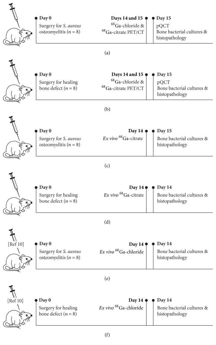
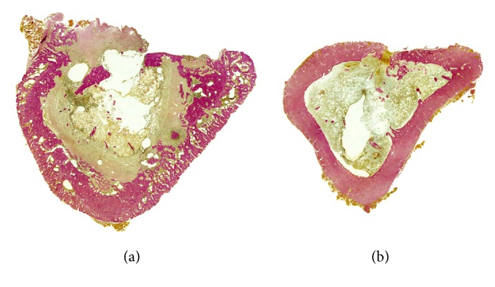
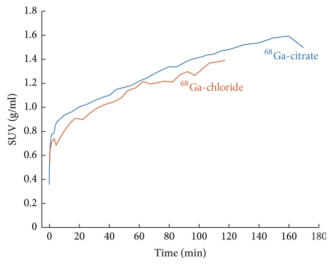
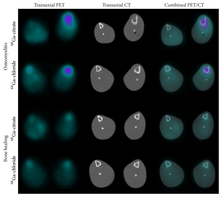
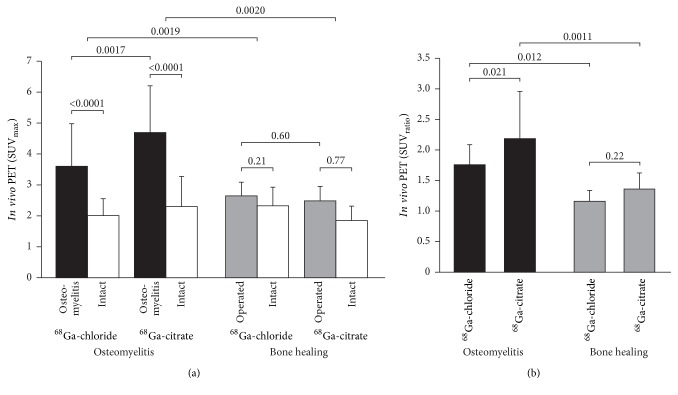
There may be some differences in the in vivo behavior of 68Ga-chloride and 68Ga-citrate leading to different accumulation profiles. This study compared 68Ga-citrate and 68Ga-chloride PET/CT imaging under standardized experimental models. Methods. Diffuse Staphylococcus aureus tibial osteomyelitis and uncomplicated bone healing rat models were used (n = 32). Two weeks after surgery, PET/CT imaging was performed on consecutive days using 68Ga-citrate or 68Ga-chloride, and tissue accumulation was confirmed by ex vivo analysis. In addition, peripheral quantitative computed tomography and conventional radiography were performed. Osteomyelitis was verified by microbiological analysis and specimens were also processed for histomorphometry. Results. In PET/CT imaging, the SUVmax of 68Ga-chloride and 68Ga-citrate in the osteomyelitic tibias (3.6 ± 1.4 and 4.7 ± 1.5, resp.) were significantly higher (P = 0.0019 and P = 0.0020, resp.) than in the uncomplicated bone healing (2.7 ± 0.44 and 2.5 ± 0.49, resp.). In osteomyelitic tibias, the SUVmax of 68Ga-citrate was significantly higher than the uptake of 68Ga-chloride (P = 0.0017). In animals with uncomplicated bone healing, no difference in the SUVmax of 68Ga-chloride or 68Ga-citrate was seen in the operated tibias. Conclusions. This study further corroborates the use of 68Ga-citrate for PET imaging of osteomyelitis.


Figures

Figure 1
Figure 1
Schematic illustration of experimental design and timing of performed analysis methods.
Figure 2
Figure 2
Histological sections of osteomyelitic (a) and control (b) rat tibias at 2 weeks after surgery. The osteomyelitic changes were characterised by a wide circumferential periosteal reaction, focally enlarged haversian canals filled with fragmented polymorphonuclear leukocytes and occasional microabscesses, and major infiltration of the bone marrow by polymorphonuclear leukocytes. In some cases, a devitalized bone fragment was seen in the unhealed cortical window. In the control animals, periosteal reaction was minimal and there was modest endosteal new bone close to the cortical defect, indicating healing of the cortical defect. Modified van Gieson stain at ×10 magnification.
Figure 3
Figure 3
Time-activity curves for 68Ga-citrate and 68Ga-chloride accumulation at the site of induced osteomyelitis in rat tibia as determined by in vivo PET/CT imaging. The line represents the mean value of two animals. The radioactivity concentration, expressed in SUV, has been decay-corrected to the time of injection.
Figure 4
Figure 4
Representative transaxial PET, CT, and combined PET/CT images of 68Ga-citrate and 68Ga-chloride accumulation at the site of induced osteomyelitis and healing bone-defects at 2 weeks after surgery. In each animal, the left tibia (on the right) underwent surgery for induction of infection or the creation of a surgical defect to represent uncomplicated bone healing, with the contralateral intact bone (on the left) serving as the control.
Figure 5
Figure 5
Quantification of 68Ga-citrate and 68Ga-chloride PET/CT imaging at 2 weeks after surgery. Bar graphs represent mean SUVmax values (±SD) (a) and SUVmax ratios, that is, operated bone-to-intact bone (b) (n = 8).

References

    1. Dinh M. T., Abad C. L., Safdar N. Diagnostic accuracy of the physical examination and imaging tests for osteomyelitis underlying diabetic foot ulcers: Meta-analysis. Clinical Infectious Diseases. 2008;47(4):519–527. doi: 10.1086/590011. - DOI - PMC - PubMed
    1. Prandini N., Lazzeri E., Rossi B., Erba P., Parisella M. G., Signore A. Nuclear medicine imaging of bone infections. Nuclear Medicine Communications. 2006;27(8):633–644. doi: 10.1097/00006231-200608000-00006. - DOI - PubMed
    1. Termaat M. F., Raijmakers P. G. H. M., Scholten H. J., Barker F. C., Patka P., Haarman H. J. T. M. The accuracy of diagnostic imaging for the assessment of chronic osteomyelitis: a systematic review and meta-analysis. The Journal of Bone and Joint Surgery—American Volume. 2005;87(11):2464–2471. doi: 10.2106/jbjs.d.02691. - DOI - PubMed
    1. Chacko T. K., Zhuang H., Nakhoda K. Z., Moussavian B., Alavi A. Applications of fluorodeoxyglucose positron emission tomography in the diagnosis of infection. Nuclear Medicine Communications. 2003;24(6):615–624. doi: 10.1097/00006231-200306000-00002. - DOI - PubMed
    1. Ledermann H. P., Kaim A., Bongartz G., Steinbrich W. Pitfalls and limitations of magnetic resonance imaging in chronic posttraumatic osteomyelitis. European Radiology. 2000;10(11):1815–1823. doi: 10.1007/s003300000480. - DOI - PubMed

Publication types

MeSH terms