. 2018 Apr 11:15:10.
doi: 10.1186/s12983-018-0259-8. eCollection 2018.

Nervous system development in the Pacific oyster, Crassostrea gigas (Mollusca: Bivalvia)

Affiliations

Nervous system development in the Pacific oyster, Crassostrea gigas (Mollusca: Bivalvia)

Olga V Yurchenko et al. Front Zool. .

Abstract

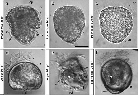
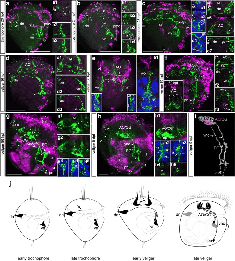
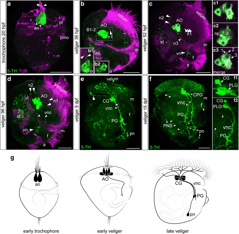
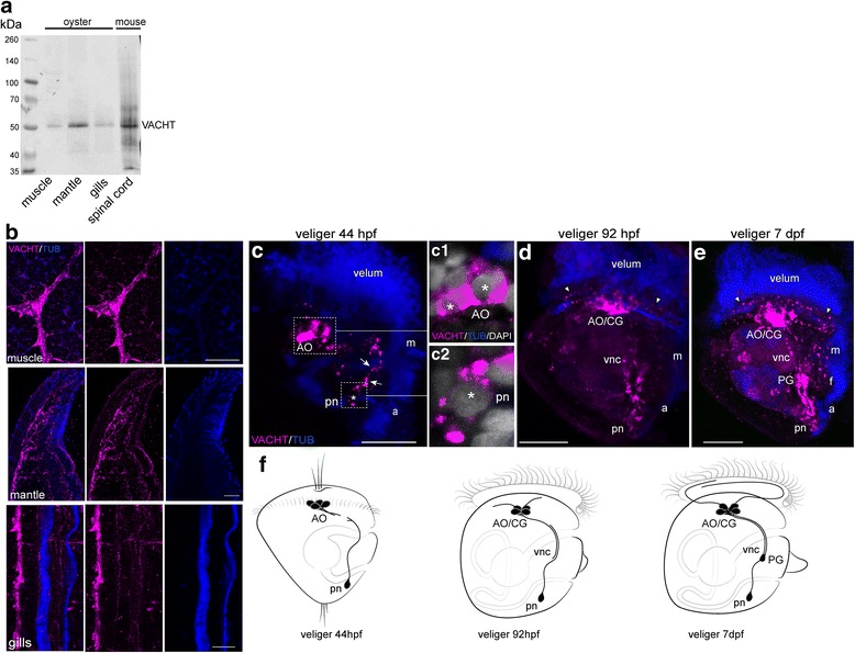
Background: Bivalves comprise a large, highly diverse taxon of invertebrate species. Developmental studies of neurogenesis among species of Bivalvia are limited. Due to a lack of neurogenesis information, it is difficult to infer a ground pattern for Bivalvia. To provide more comprehensive morphogenetic data on bivalve molluscs and relationships among molluscan clades, we investigated neurogenesis in the Pacific oyster, Crassostrea gigas, from the appearance of the first sensory cells to the formation of the larval ganglionic nervous system by co-immunocytochemistry of the neuronal markers FMRFamide or 5-HT and vesicular acetylcholine transporter (VAChT).

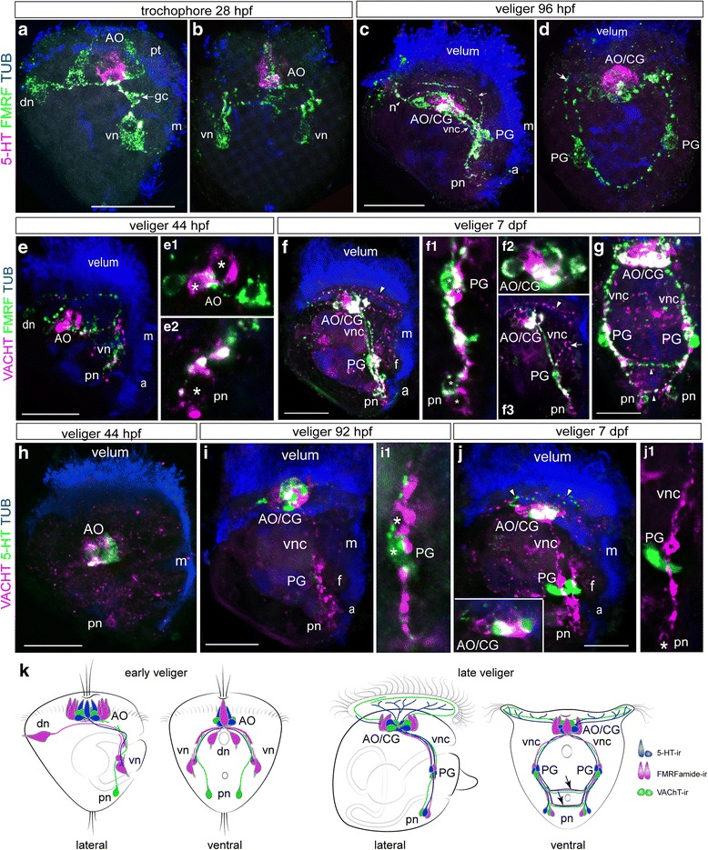
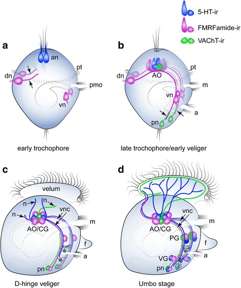
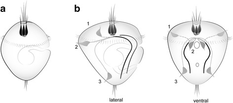
Results: Neurogenesis begins with the emergence of the apical serotonin-immunoreactive (5-HT-ir) sensory cells and paired sensory posttrochal dorsal and ventral FMRFamide-immunoreactive (FMRFamide-ir) cells at the early trochophore stage. Later, at the early veliger stage, the apical organ (AO) includes 5-HT-ir, FMRFamide-ir, and VAChT-ir cells. At the same stage, VAChT-ir cells appear in the posterior region of larvae and send axons towards the AO. Thus, FMRFamide-ir neurites and VAChT-ir processes form scaffolds for longitudinal neurite bundles develop into the paired ventral nerve cords (VNC). Later-appearing axons from the AO/CG neurons join the neurite bundles comprising the VNC. All larval ganglia appear along the VNC as paired or fused (epiathroid) clusters in late veliger and pediveliger larvae. We observed the transformation of the AO into the cerebral ganglia, which abundantly innervated the velum, and the transformation of ventral neurons into the pedal ganglia, innervating the foot, gills, and anterior adductor muscle. The visceral ganglia appear last in the pediveliger oyster and innervate the visceral mass and posterior adductor of premetamorphic larvae. In addition, a local FMRFamide-ir network was detected in the digestive system of pediveliger larvae. We identified VAChT-ir nervous elements in oyster larvae, which have not been observed previously in molluscs. Finally, we performed a morphology-based comparative analysis of neuronal structures among bivalve, conchiferan, and aculiferan species.

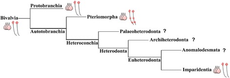
Conclusions: We described the development of the nervous system during the larval development in Crassostrea gigas. These data greatly advance the currently limited understanding of neurodevelopment in bivalves and mollusks, which has hampered the generation of a ground pattern reconstruction of the last common ancestor of Mollusca. Our morphological data support phylogenomic data indicating a closer Bivalvia-Gastropoda sister group relationship than the Bivalvia-Scaphopoda (Diasoma) group relationship.

Keywords: Acetylcholine; Evolution; FMRFamide; Larvae; Mollusca; Neurogenesis; Serotonin.


Conflict of interest statement

The field studies did not involve endangered or rare invertebrate species. No specific permissions were required to access the marine area, as it falls within Russian state-owned land.All authors agree on the submission and publication of this paper and its included figures and tables.The authors declare that they have no competing interests.Springer Nature remains neutral with regard to jurisdictional claims in published maps and institutional affiliations.

Figures

Fig. 1
Fig. 1
Stages of Crassostrea gigas larval development. Light microscopy images. Early (a), middle (b), and late (c) trochophores and early (D-hinge, d), middle (e), and pedi-umbo-stage (f) veligers. Abbreviations: ad - anterior adductor, ep – episphere, es – esophagus, hp. – hyposphere, in – intestine, m – mouth, pmo – presumptive mouth opening, pt. – prototroch, ret. – retractors, s – shell, sg – shell gland, st – stomach, tt – telotroch, u – umbo, v – velum. Scale bars = 20 μm
Fig. 2
Fig. 2
FMRFamide-immunoreactivity (FMRFa-ir) in the trochophore and veliger larvae of C. gigas. Green—FMRFa-ir; magenta—cilia, acetylated tubulin immunoreactivity; blue—nuclei, DAPI. The apical pole is always upward; the ventral side is on the right. a Trochophore at 20 hpf. Dorsal (arrowheads) and ventral (asterisks) groups of early neurons located posttrochally. a1 Higher magnification of right dorsal neuron (dn); arrowheads indicate a ciliated dendritic knob (ck) and basal neurites (n). a2 and a3 Right and left ventral neurons (vn, asterisks), respectively. b Trochophore at 24 hpf. Faint dotted immunoreactivity appears at the apical region (arrows). A dorsal cell sends an axon towards the ventral side (arrowheads). Three ventral cells indicated by one arrow (flask-shaped cells) and asterisks (round cells). b1 and b2 Higher magnification of the right and left dorsal neurons (dn), arrowheads indicate the ciliated dendritic knob (ck) of each cell. b3 and b4 Higher magnification of right and left ventral groups (vn); each ventral group contains two flask-shaped cells (arrows) and two round cells (asterisks). c Trochophore at 28 hpf. Two flask-shaped cells appear in the apical organ (AO). Growth cone (arrows, gc) of the right dorsal cell (dn), where a growing axon reaches the ipsilateral ventral cell (vn); only the right sides of the larvae are shown. c1-c4 Right and left dorsal and ventral groups of neurons with DAPI. c5 and c6 Two flask-shaped neurons of the AO at two different magnifications (asterisks). c7 Higher magnification of a growing axon of the right dorsal cell; arrowhead indicates a ciliated dendritic knob, and arrows indicate a growth cone of the dorsal neuron. d D-hinge veliger stage at 36 hpf. The cells of the AO are located at the top (asterisks); no connections exist between the AO and other early cells. d1 Two flask-shaped apical neurons (asterisks) and their basal neurites. d2 and d3 Long axons of both the right and left dorsal cells reach the ipsilateral groups of the ventral cells. e Ventral view of the veliger at 36 hpf. Arrowheads indicate projections from dorsal and apical cells towards the ventral neurons. insets Higher magnification of the left and right ventral groups. e1 Higher magnification of the apical neurons demonstrating long cilia at the end of their dendrite (arrows). f The veliger stage at 52 hpf and 60 hpf (insert). Dorsal (dn) and ventral (vn) cells stain in a punctate pattern and their axons form an anlagen of the ventral nerve cord. Small immunopositive neurons appear posteriorly (posterior neurons, arrow, pn). Insert: Four flask-shaped apical neurons (asterisks) of AO.f1 Higher magnification of the AO (asterisk). f2 Long axon of a dorsal neuron (dn) to the ventral neuron (vn). f3 Neurites extending from the AO (arrow, an) follow the path established by pioneer axons of the dorsal neurons. g The veliger stage at 96 hpf. AO/CG consist of six flask-shaped cells (asterisks) and basal processes of AO/CG neurons organize into a compact central neuropil (arrow, np). A single neurite (n, arrowhead) extends from the neuropil of the AO/CG toward the velum. Paired ventral cords (vnc) with the interconnecting commissures (short arrows) are clearly visible. The anlagen of the pedal ganglia (PG) appear along each ventral cord in the region of the developing foot. g1 Flask-shaped neurons (asterisks) with neuropil forming (Z12). g2 Flask-shaped neurons (asterisks) with neuropil forming (Z14). g3 and g4 Neurons within the PG under high magnification (asterisks). g5 Posterior neurons under high magnification (asterisk). h The late veliger stage at 9 dpf. Neurites extend from the AO/CG to the dorsal side and dorsal edge of the velum (arrowheads). The anlagen of the PG are located along the ventral cord (vnc). Paired ventral cords are connected by two commissures (short arrows). h1 Compact neuropil in the center of the AO/CG. h2 and h3 Right and left PG anlagen; arrows point to the neuronal nuclei. h4 and h5 Higher magnification of the right and left posterior neurons; the asterisk marks the cell nucleus. i 3D reconstruction of the AO/CG complex, paired ventral cords and PG anlagen of 9-dpf late-veliger larvae. j Summary diagram of the ontogeny of the FMRFa-ir-containing structures in C. gigas. Only one lateral side is shown. Arrowhead indicates an axon of a dorsal cell in the late trochophore. Additional abbreviations: a – anus, f – foot, m – mouth, pmo – presumptive mouth opening, pt – prototroch, st – stomach. Scale bars =20 μm
Fig. 3
Fig. 3
Serotonin immunoreactivity (5-HT-ir) in the trochophore and veliger larvae of Crassostrea gigas. Green—5-HT-ir; magenta—cilia, acetylated tubulin immunoreactivity. The apical pole is always upward; the ventral side is on the right. a The early trochophore at 20 hpf. The first two flask-shaped neurons (arrows, an) are located in the anterior extreme of the larval body. Each cell has a ciliated short dendrite (arrowheads). b D-hinge veliger at 36 hpf. 5-HT-ir cells comprise a compact apical organ (AO) and comprise flask-shaped and round cells each with thin neurites (arrowheads, n1) running to the velum. b1 two flask-shaped cells with thin neurites and neuropil (np). b2 single round cells (black asterisk). c The veliger stage at 52 hpf. Cells of the AO extend three long neurites to the velum (arrowheads, n1), anterior−dorsal (arrowheads, n2), and posterior−ventral (arrowheads, n3). Acetylated tubulin-ir reveals the digestive system and green autofluorescent particles are visible in the stomach (st) in b, c, d, e. insets: High magnification of the AO cell composition. c1 two flask-shaped and one round cells (black asterisk) (Z5), c2 third flask-shaped cells and second round cell (black asterisk), and neuropil (arrows, np) c3 merge picture. There are five cells: three flask-shaped cells and two round cells (black asterisk); d The middle veliger stage at 96 hpf. Two posterior−ventral neurites from cells of the AO run in parallel along the ventral part of the larval body organizing the ventral nerve cords (vnc). e The veliger at 9 dpf. Cerebral ganglia (CG) are located at the top, and pedal ganglia (PG) are detected in the middle of the ventral nerve cord. Small immunoreactive posterior neurons (pn) are located near the caudal end of each ventral cord. Neurites extending from the CG have multiple branches in the velum region. Note the solitary branch extending from the apical part of the ganglion (arrowhead) towards the dorsal edge of the velum. f The veliger at 15 dpf. Note the paired ventral nerve cords with two commissures (short arrows). Cerebral ganglia located on top of the ventral cords. The thickening of the upper portion of each ventral cord represents the anlagen of the pleural ganglia (PLG) and together with the CG they form a fused CG/PLG complex indicated as CPG. Right and left PG are located along the respective ventral cords at the middle of the foot. Posterior neurons are located caudally, and each sends an axon to the ipsilateral ventral cord. The velum is richly innervated by fibers extending from the CPG. Thin neurites extend to the foot, and digestive organs (mark as peripheral nerve system, PNS) extend from the PG region (arrowheads). insets: f1 High magnification of the CG/PLG region; f1 focus on CG and PLG parts of CPG, PG, ventral cord, and peripheral innervation. g Summary diagram of the ontogeny of the 5-HT-ir-containing structures in C. gigas. Only one lateral side is shown. Additional abbreviations: f – foot, m – mouth, pmo – presumptive mouth opening, pt. – prototroch, st – stomach. Scale bars =20 μm
Fig. 4
Fig. 4
Characterization of VAChT antibodies and VAChT-ir (VAChT, magenta) in oyster, Crassostrea gigas, tissues. a Western blot of total protein lysates from adult oyster tissue probes stained with goat polyclonal antibodies against rat VAChT. The specific band is detected in all tested oyster tissues as well as in cell lysate from the mouse spinal cord. b Double-staining for VAChT/tubulin (VAChT/TUB) of frozen sections of adult oyster tissues. A strong positive signal is detected in the anterior adductor muscle (muscle), mantle, and gills at the structures corresponding to the nerve bundles. c-e Confocal images of the larvae stained with VAChT/tubulin, right side view; the anterior is always up. c D-hinge veliger. The apical organ (AO) contains two to three cells and their basal fibers. Paired solitary neurons located posteriorly on the right and left sides of the larval body (pn). Each posterior cell sends an anteriorly directed fiber along the ventral edge of the larval body (arrows) (an asterisk marks the neuron nuclei). c1 Bodies of two AO neurons; asterisks mark the nuclei. c2 Posterior neuron, an asterisk marks the nucleus. d In the veliger stage (92 hpf), a strong VAChT-ir signal is detected within the AO/CG complex, and single fibers appear to innervate the velum (arrowheads). Immunopositive fibers run in two parallel, ventral cords (vnc), and a solitary posterior cell is visible at the caudal end of each ventral cord. Immunopositive cells appear in the PG. e At the 7-dpf veliger stage, VAChT-ir fibers from AO/CG innervate the velum, and fibers from PG innervate the foot (f) and regions around the mouth. g Summary diagram of the ontogeny of the VAChT-ir-containing structures in C. gigas. Only one lateral side is shown. Additional abbreviations: a – anus, m – mouth. Scale bars =100 μm in b and 20 μm in c
Fig. 5
Fig. 5
Double staining of 5-HT/FMRFa-ir, VAChT/FMRFa-ir, and VAChT/5-HT-ir nervous elements combined with tubulin immunoreactivity of ciliary structures in Crassostrea gigas larvae. Neurotransmitter co-localization appears white in all pictures. a and b Side and front views, respectively, of the alternative expression of FMRFa-ir and 5-HT-ir within early neurons at the 28-hpf trochophore stage. In the apical organ (AO), 5-HT-ir neurons are surrounded by FMRFa-ir neurons. Arrows point to the growth cone of the right dorsal cell (gc). c and d Alternative expression of FMRFa-ir and 5-HT-ir in cells of the AO/cerebral ganglia complex (AO/CG) at the 96-hpf stage. Only FMRFa-ir cells are present in the pedal ganglia (PG) anlagen. Arrows point to the peripheral process from the PG to the velum, and an arrowhead marks the process (n) from the AO/CG to the velum. e Alternative expression of VAChT-ir and FMRFa-ir within the early neurons (dn, vn, pn, and AO) in the 44-hpf veliger. e1 VAChT-ir cells (asterisks) are surrounded by FMRFa-ir cells in the AO. e2 A posterior VAChT-ir cell (pn) sends a process along the FMRFa-ir fiber; an asterisk marks the posterior cell nucleus. f and g Side and front views in the 7-dpf veliger stage demonstrate the presence of both VAChT-ir and FMRFa-ir elements in the AO/CG complex, PG, and posterior neurons (pn). Partial co-localization occurs in the AO/CG neuropil and in the ventral nerve cords. f1 and f2 Micrograph demonstrating alternative VAChT-ir and FMRFa-ir expression in PG and AO/CG neurons, their partial co-localization within the AO/CG and the PG neuropil, and the processes of the ventral nerve cords (vnc). f3 A VAChT-ir process runs from the AO/CG to the velum (arrowhead) and from the PG to the foot anlagen (arrow). h Alternative expression of VAChT-ir and 5-HT in the 44-hpf veliger stage. The AO contains both VAChT-ir and 5-HT-ir neurons, while a posterior neuron (pn) exhibits VAChT-ir only. i In the 92-hpf veliger stage, VAChT and 5-HT are alternatively seen within the cell bodies of the AO/CG and are partly co-localized in the neuropil. i1 Magnification of 5-HT-ir and VAChT-ir neurons in PG. A 5-HT-ir process runs along the VAChT-ir fiber; asterisks mark the cell bodies. j In the 7-dpf veliger stage, both VAChT-ir and 5-HT-ir fibers emanating from the AO/CG run to the velum (arrowheads). inset: Alternative expression of VAChT-ir and 5-HT-ir within the AO/CG cell bodies and their partial co-localization within the neuropil. j1 5-HT-ir within the PG appears to be adjacent to a VAChT-ir nerve bundle in the ventral nerve cord. An asterisk marks the body of the posterior VAChT-ir cell (pn). k Summary diagram of the ontogeny of the VAChT-ir, 5-HT-ir, and FMRFamide-ir-containing structures in C. gigas. Only one lateral side is shown. Additional abbreviations: a – anus, m – mouth, pt. – prototroph. Scale bars =20 μm
Fig. 6
Fig. 6
FMRFamide-immunoreactivity (FMRFa-ir) combined with phalloidin staining and serotonin-immunoreactivity (5-HT-ir) in the pediveliger stage (28 dpf) of Crassostrea gigas. Magenta—muscles, phalloidin; blue—nuclei, DAPI. The apical pole is always upward, and the ventral side is on the right. a General view of FMRFa-ir in the 28-dpf pediveliger stage with the cerebro-pleural ganglion (CPG), paired ventral cords (vnc), pedal (PG), and ventral (VG) ganglia located along the vnc. Note the absence of special cerebro-pedal cords separated from the vnc bundles. Solitary posterior neurons (pn) are visible at the most caudal part of each vnc. Peripheral innervations of the velum, gill rudiment (gr), anterior and posterior adductor muscles (an.ad. and post.ad.) and body wall are visible. Inset: High magnification of the PG/VG region exhibiting a chain of FMRFa-ir cells within the VG rudiment. a1 Innervation of anterior adductor muscle (an.ad.) with fibers emanating from the PG. Retractor muscles (ret) are innervated from the VG. a2 A network of thin FMRFa-ir fibers in the foot (f). a3 The FMRFa-ir network along the digestive system (arrowheads, dig.s.) forms a network of the enteric nerve system (ENS). The posterior adductor (post.ad) is innervated by a thick fiber from the VG. inset: High magnification view of the developing gill rudiment (gr) with two parallel, branched fibers emanating from the PG. b General view of 5-HT-ir in the 28-dpf pediveliger stage with CPG, pedal (PG), and ventral (VG) ganglia located along the ventral nerve cords. Solitary posterior neurons (pn) are visible near the most caudal part of each vnc. Peripheral innervations of the velum and anterior and posterior adductor muscles (an.ad. and post.ad.) are visible. c General view of 5-HT in the 35-dpf pediveliger with a well-developed cerebro-pleural ganglion (CPG), and pedal (PG) and ventral (VG) ganglia. Inset: High magnification of the CPG. c1 The anterior adductor and velum are innervated from the CPG and PG. The posterior adductor is innervated from the ventral ganglia. Scale bars =50 μm
Fig. 7
Fig. 7
Phylogenic tree of major bivalve lineages based on González et al. [1], revealing ground patterns based on morphological data of neurogenesis of bivalves that have been investigated. Pink cells are 5-HT-ir cells of the AO in the studied species. Ventral nerve cord (VNC, black parallel lines) with red ganglia (top is the cerebral, middle is the pedal, and bottom is the ventral ganglion). Protobranchia (non-published data), Pteriomorpha, and Imparidentia show three flask-shaped cells in the AO. Within Pteriomorpha mussels and oysters possess a VNC with three paired ganglia, while Protobranchia and Imparidentia have a VNC with only a cerebral ganglion
Fig. 8
Fig. 8
Summary schematic of the early neurogenetic events of the Pacific oyster, Crassostrea gigas. Serotonin-immunoreactive (5-HT-ir) elements are blue; FMRFa-ir elements are violet; and VAChT-immunoreactive (VAChT-ir) elements are green. The apical pole is always upward, and the ventral side is on the right. a At the early trochophore stage, only 5-HT-ir cells are present in the apical region of the episphere. Apical neurons (an) have a flask shape and bear cilia. Paired dorsal (dn) and ventral neurons (vn) located in the hyposphere region express FMRFa-ir. The dorsal cells have a ciliated sensory dendrite and an axon extending towards the ventral cells (arrowheads); VAChT-ir elements are absent. b By the late trochophore/early veliger stage, the axons of the dorsal cells pass a group of ventral cells to reach the caudal region of the larvae. VAChT-ir neurons appear posteriorly (pn) and send axons anteriorly along the FMRFa-ir processes of the dorsal cells. Thus, the paired ventral nerve cords are established by early FMRFa-ir and VAChT-ir nerve elements. FMRFa-ir and VAChT-ir flask-shaped cells are incorporated into the apical organ (AO), and AO neurons are located between the ventral cords but have no connections with them. c In the D-hinge veliger stage, solitary FMRFa-ir neurons (pn) appear posteriorly near the posterior VAChT-ir cells. Axons of apical, ventral, and posterior neurons grow along the ventral cords. Two commissures (asterisks) connect the left and right ventral cords (vnc). The AO is located between the two cerebral ganglia forming the AO/CG-complex. 5-HT-ir, VAChT-ir, and FMRFa-ir neurites (arrowheads, n) extend from the AO/CG neuropil. d In the umbo stage, the pedal (PG) and visceral ganglia (VG) adjoin the longitudinal ventral cords. No specific cerebro-pedal cords can be separated within the bands of the ventral cords. The CPG, PG, and posterior group contain FMRFa-ir, 5-HT-ir, and VAChT-ir neurons, while the VG only contains FMRFa-ir and 5-HT-ir cells. Both ventral cords and commissures contain FMRFa-ir, 5-HT-ir, and VAChT-ir neurites. 5-HT-ir and VAChT-ir processes extending from the CG richly innervate the ciliated velum. 5-HT-ir and FMRFa-ir neurites innervate the velum anterior edge. Abbreviations: a – anus, an – apical neuron, CG – cerebral ganglia, dn – dorsal neurons, m – mouth, n – peripheral neurites, PG – pedal ganglia, pmo – presumptive mouth opening, pt. – prototroch, VG – visceral ganglia, vn – ventral neurons, vnc – ventral nerve cords
Fig. 9
Fig. 9
Schematic presentation of the ground structures of the nervous system in Bivalvia and Mollusca based on morphological data of neurogenesis in studied species. a suggested ground pattern of AO of the last common ancestor of Bivalvia consist of three flask-shaped cells. b the most conserved nervous structures in Mollusca are an AO consisting of three flask-shaped cells and a paired ventral nerve cord (black lines). Note, the neurites of pioneer peripheral sensory cells are located in the episphere (1st scenario for Mytillus trossulus, Bivalvia) or hyposphere (located dorsally (2nd scenario for oyster) or caudally (3rd scenario for chitons and gastropods); all trace the future VNC. Thus, the position of first sensory cells in larvae does not matter, but the route that the neurites make is necessary for positioning of the VNC later

References

    1. González VL, Andrade SCS, Bieler R, Collins TM, Dunn CW, Mikkelsen PM, Taylor JD, Giribet G. A phylogenetic backbone for Bivalvia: an RNA-seq approach. Proc R Soc B. 2015;282:20142332. doi: 10.1098/rspb.2014.2332. - DOI - PMC - PubMed
    1. Dame RF. Ecology of marine bivalves: an ecosystem approach. 2nd ed. New York: CRC press; 2011.
    1. Raineri M, Ospovat M. The initial development of gangliar rudiments in a posterior position in Mytilus galloprovincialis (Mollusca: Bivalvia) J Mar Biol Assoc UK. 1994;74:73–77. doi: 10.1017/S0025315400035670. - DOI
    1. Croll RP, Jackson DL, Voronezhskaya EE. Catecholamine-containing cells in larval and post larval bivalve molluscs. Biol Bull. 1997;193:116–124. doi: 10.2307/1542757. - DOI - PubMed
    1. Voronezhskaya EE, Nezlin LP, Odintsova NA, Plummer JT, Croll RP. Neuronal development in larval mussel Mytilus trossulus (Mollusca; Bivalvia) Zoomorphology. 2008;127:97–110. doi: 10.1007/s00435-007-0055-z. - DOI

LinkOut - more resources