. 2018 Apr 12:10:31.
doi: 10.1186/s13098-018-0332-7. eCollection 2018.

Possible neuroprotective role of P2X2 in the retina of diabetic rats

Affiliations

Possible neuroprotective role of P2X2 in the retina of diabetic rats

Jorge E Mancini et al. Diabetol Metab Syndr. .

Abstract

Background: Purinergic receptors are expressed in different tissues including the retina. These receptors are involved in processes like cell growth, proliferation, activation and survival. ATP is the major activator of P2 receptors. In diabetes, there is a constant ATP production and this rise of ATP leads to a persistent activation of purinergic receptors. Antagonists of these receptors are used to evaluate their inhibition effects. Recently, the P2X2 has been reported to have a neuroprotective role.

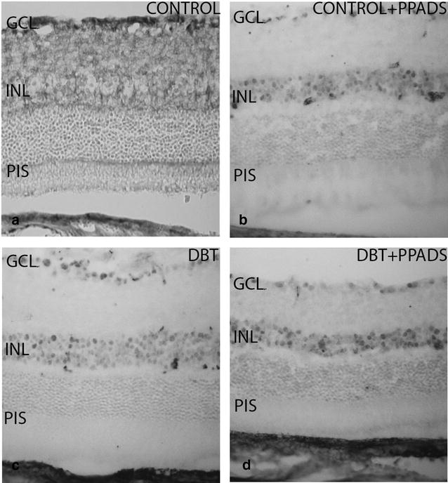
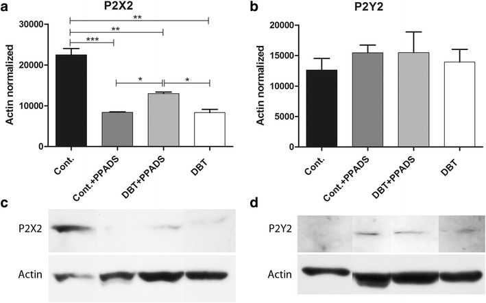
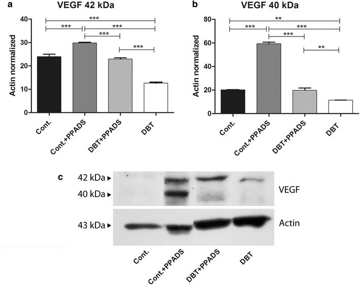
Methods: We carried out a study in groups of diabetic and non-diabetic rats (N = 5) treated with intraperitoneal injections of PPADS, at 9 and 24 weeks of diabetes. Control group received only the buffer. Animals were euthanized at 34 weeks of diabetes or at a matching age. Rat retinas were analyzed with immunohistochemistry and western blot using antibodies against GFAP, P2X2, P2Y2 and VEGF-A.

Results: Diabetic animals treated with PPADS disclosed a much more extended staining of VEGF-A than diabetics without treatment. A lower protein expression of VEGF-A was found at the retina of diabetic animals without treatment of purinergic antagonists compared to diabetics with the antagonist treatment. Inhibition of P2X2 receptor by PPADS decreases cell death in the diabetic rat retina.

Conclusion: Results might be useful for better understanding the pathophysiology of diabetic retinopathy.

Keywords: Diabetic retinopathy; P2X2; PPADS; Retina; VEGF-A.


Figures

Fig. 1
Fig. 1
Fast glycemia. The two bar charts show the analyses of fast glycemia in two different periods in the seven groups of animals. a Glycemia levels at 9 weeks after vehicle or streptozotocin treatment in control and diabetic animals. Measurement was done 1 day before PPADS first cycle treatment. All control groups had normal glycemia levels before purinergic receptors antagonist treatment. b Glycemia levels at 34 weeks after two purinergic receptors antagonist treatment cycles performed at 9 and 24 weeks of diabetes. Cont. control; PPADS pyridoxalphosphate-6-azophenyl-2′,4′-disulfonic acid; DBT diabetic
Fig. 2
Fig. 2
Retinal cross section. Immunofluorescent analyses of diabetic and control animals without purinergic receptors antagonist treatment. A Diabetic group (34 weeks after STZ IP injection). B Control group. P2Y2r immunoreactivity was seen in the fiber layer of diabetic and control animals (arrow-A1 and B1). In the diabetic group staining had a larger extension. Positive immunoreactivity of GFAP was observed in glial cells of diabetics and controls (arrow A2 and B2). In diabetic animals immunoreactivity was much more extended and some GFAP positive cells showed a morphology similar to that seen in Müller Cells (arrow head-A2). Co-expression of P2Y2 and GFAP in diabetics and controls had different patterns. In diabetic rats co-expression of P2Y2 and GFAP was seen in the fiber layer and in glial cells extending to the photoreceptor layer (A3), while in controls the co-expression was slightly observed in the fiber layer (B3)
Fig. 3
Fig. 3
Retinal cross-sections. P2Y2 immunohistochemistry in diabetic and control animals treated with purinergic receptors antagonist (PPADS). a and b represent control groups. c and d represent diabetic groups. b and d groups were treated with intraperitoneal injection of group with PPADS. P2Y2r immunostaining was seen in the photoreceptor inner segment in all diabetics and in controls treated with purinergic receptors antagonist. GCL ganglion cells layer; INL Inner nuclear layer; PIS photoreceptor inner segment; DBT diabetic; PPADS animals treated with pyridoxalphosphate-6-azophenyl-2′,4′-disulfonic acid
Fig. 4
Fig. 4
Retinal cross-sections. P2X2 immunohistochemistry in diabetic and control animals treated with purinergic receptors antagonist (PPADS). a and b Represent control groups. c and d Represent diabetic groups. b and d Groups were treated with intraperitoneal injection of PPADS. P2X2r immunostaining was seen in the photoreceptor inner segment and outer nuclear layer of controls without treatment. In the remaining animal groups immunoreactivity was found in the inner nuclear layer. GCL ganglion cells layer; INL Inner nuclear layer; PIS photoreceptor inner segment; DBT diabetic; PPADS animals treated with pyridoxalphosphate-6-azophenyl-2′,4′-disulfonic acid
Fig. 5
Fig. 5
Immunohistochemical analyses of VEGF-A. Immunostaining of VEGF-A was found in two different patterns. Controls with PPADS treatments showed a positive immunoreactivity in GCL, INL and PIS. Controls without treatment and c, d diabetic animals showed staining in structures which may be small vessels (arrows). GCL ganglion cells layer; INL Inner nuclear layer; PIS photoreceptor inner segment; DBT diabetic; PPADS animals treated with pyridoxalphosphate-6-azophenyl-2′,4′-disulfonic acid
Fig. 6
Fig. 6
Western blot analyses of P2X2 and P2Y2. a P2X2 densitometry quantification, lower expression was seen in the diabetic and treated groups. b P2Y2 densitometry quantification, non-significant differences were found between groups. c P2X2 immunoblot. d P2Y2 immunoblot. Diabetic animals treated with PPADS showed higher P2X2 expression levels compared with non-treated diabetic group. Cont. control; PPADS pyridoxalphosphate-6-azophenyl-2′,4′-disulfonic acid; DBT diabetic (*p<0.05; **p<0.01; ***p<0.001)
Fig. 7
Fig. 7
Western blot analyses of VEGF-A. a 42 KDa band. The expression was lower in diabetic group compared to control group or PPADS groups. b 40 KDa band. The expression profile was similar than 42 kDa band. c Inmunoblot. Cont. control; PPADS pyridoxalphosphate-6-azophenyl-2′,4′-disulfonic acid; DBT diabetic (*p<0.05,**p<0.01, ***p<0.01)
Fig. 8
Fig. 8
a Retinal ganglion cell counting. b Retinal thickness. c Cryosection stained with H&E for control group without treatment. d Cryosection stained with H&E for treated diabetic group. e Cryosection stained with H&E for diabetic group. Cont. control; PPADS pyridoxalphosphate-6-azophenyl-2′,4′-disulfonic acid; DBT diabetic (*p<0.05,**p<0.01, ***p<0.01)

Similar articles

Cited by

References

    1. Ralevic V, Burnstock G. Receptors for purines and pyrimidines. Pharmacol Rev. 1998;50:413–492. - PubMed
    1. Lazarowski ER, Boucher RC, Harden TK. Mechanisms of release of nucleotides and integration of their action as P2X- and P2Y-receptor activating molecules. Mol Pharmacol. 2003;64:785–795. doi: 10.1124/mol.64.4.785. - DOI - PubMed
    1. Lambrecht G, Braun K, Damer M, Ganso M, Hildebrandt C, Ullmann H, et al. Structure-activity relationships of suramin and pyridoxal-5′-phosphate derivatives as P2 receptor antagonists. Curr Pharm Des. 2002;8:2371–2399. doi: 10.2174/1381612023392973. - DOI - PubMed
    1. Fries JE, Wheeler-Schilling TH, Guenther E, Kohler K. Expression of P2Y1, P2Y2, P2Y4, and P2Y6 receptor subtypes in the rat retina. Invest Ophthalmol Vis Sci. 2004;45:3410–3417. doi: 10.1167/iovs.04-0141. - DOI - PubMed
    1. Puthussery T, Fletcher EL. P2X2 receptors on ganglion and amacrine cells in cone pathways of the rat retina. J Comp Neurol. 2006;496:595–609. doi: 10.1002/cne.20889. - DOI - PubMed

LinkOut - more resources