

Neurosurgical Interventions for Occipital Encephalocele
- PMID: 29682014
- PMCID: PMC5898085
- DOI: 10.4103/1793-5482.228549
Neurosurgical Interventions for Occipital Encephalocele
Abstract
Aims and objectives: The aim of this study is to find the outcome of repair and resection of the occipital encephalocele.
Study design: Case series.
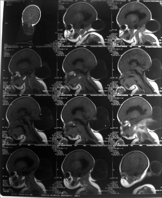
Materials and methods: The clinical data of fifty consecutive occipital encephalocele patients were retrieved from medical records including operative notes, postoperative follow-up visits, and postsurgical complications were noted for analysis from November 2009 to November 2013 at the Department of Neurosurgery, Jinnah Postgraduate Medical Centre, Karachi, Pakistan. All patients were assessed by computed tomography scan, magnetic resonance imaging brain, and ultrasound when needed. Physician's assessment, physical examination, and his/her questions to the family at follow-up were used as a tool to determine if there was a developmental delay rather than quantitative analysis like hydrocephalus questionnaires. Patients who developed complications and delayed milestone were regarded as no improvement and those who did not develop complications and achieved appropriate milestone were regarded as improved at 18 months follow-up.
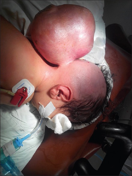
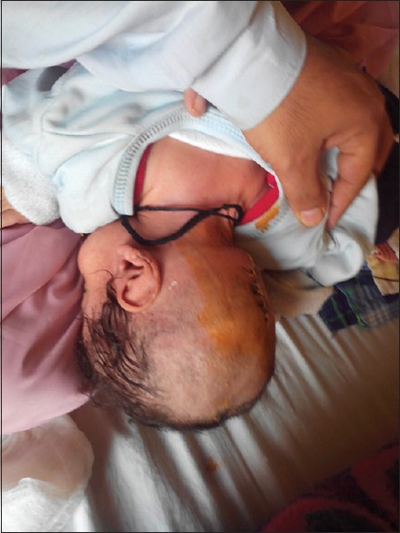
Results: Of 50 patients, 17 were males and 33 were females. The average age at presentation was 2.4 months. 16 (32%) patients had increased head circumference and hydrocephalus, 2 (4%) had associated Dandy-Walker cyst, 3 (6%) developed developmental delays, and 8 (15%) had a seizure disorder. None of our patients had neurological deficits. The size of the sac ranged from 2 cm × 3 cm to 27 cm × 15 cm. 9 (18%) patients were admitted with the complication of sac rupture and 2 (4%) patients sac ruptured after admission. Only one patient (2%) had a cerebrospinal fluid leak postoperatively that was repaired primarily without patch graft or dura seal while 4 (8%) developed hydrocephalus after repair of the sac which was treated with placement of ventriculoperitoneal shunt. One (2%) patient did not recover from anesthesia and expired.
Conclusion: Encephalocele is commonly seen in the practice of neurosurgery in the world as well as in Pakistan. Modern neuroimaging, neurosurgical techniques, and neonatal neurological intensive care have greatly improved morbidity and mortality in the care of encephalocele.
Keywords: Cerebrospinal fluid; IQ; encephalocele; hydrocephalus; ventriculoperitoneal shunt.
Conflict of interest statement
There are no conflicts of interest.
Figures
Similar articles
-
Hydrocephalus and occipital encephaloceles: presentation of a series and review of the literature.Childs Nerv Syst. 2021 Nov;37(11):3437-3445. doi: 10.1007/s00381-021-05312-7. Epub 2021 Aug 14. Childs Nerv Syst. 2021. PMID: 34390379 Review.
-
Giant Encephalocele in Sokoto, Nigeria: A 5-Year Review of Operated Cases.World Neurosurg. 2020 Jul;139:51-56. doi: 10.1016/j.wneu.2020.03.061. Epub 2020 Mar 19. World Neurosurg. 2020. PMID: 32201291
-
Risk factors for hydrocephalus and neurological deficit in children born with an encephalocele.J Neurosurg Pediatr. 2015 Apr;15(4):392-8. doi: 10.3171/2014.10.PEDS14192. Epub 2015 Feb 6. J Neurosurg Pediatr. 2015. PMID: 25658248 Free PMC article.
-
Frontal and occipital horn ratio is associated with multifocal intraparenchymal hemorrhages in neonatal shunted hydrocephalus.J Neurosurg Pediatr. 2017 Nov;20(5):432-438. doi: 10.3171/2017.6.PEDS16481. Epub 2017 Sep 8. J Neurosurg Pediatr. 2017. PMID: 28885094
-
Occipital encephalocele associated with Dandy-Walker malformation: a case-based review.Childs Nerv Syst. 2022 Sep;38(9):1683-1688. doi: 10.1007/s00381-022-05551-2. Epub 2022 May 19. Childs Nerv Syst. 2022. PMID: 35588332 Review.
Cited by
-
Giant occipital encephalocele complicated with obstructive hydrocephalus: A case report.Ann Med Surg (Lond). 2022 Jul 12;80:104176. doi: 10.1016/j.amsu.2022.104176. eCollection 2022 Aug. Ann Med Surg (Lond). 2022. PMID: 35855875 Free PMC article.
-
Occipital encephalocele: Presentation of case.Int J Surg Case Rep. 2023 Sep;110:108642. doi: 10.1016/j.ijscr.2023.108642. Epub 2023 Aug 9. Int J Surg Case Rep. 2023. PMID: 37651806 Free PMC article.
-
Hydrocephalus and occipital encephaloceles: presentation of a series and review of the literature.Childs Nerv Syst. 2021 Nov;37(11):3437-3445. doi: 10.1007/s00381-021-05312-7. Epub 2021 Aug 14. Childs Nerv Syst. 2021. PMID: 34390379 Review.
-
Posterior vault encephaloceles: from antenatal management to post-surgical follow-up-a cooperative study.Childs Nerv Syst. 2025 Feb 24;41(1):121. doi: 10.1007/s00381-025-06764-x. Childs Nerv Syst. 2025. PMID: 39994065 Free PMC article.
-
Occipital encephalocele in a neonate: a case successfully managed by excision and formation of a reverse visor scalp flap.BMJ Case Rep. 2020 Jan 21;13(1):1-4. doi: 10.1136/bcr-2019-232127. BMJ Case Rep. 2020. PMID: 31969420 Free PMC article.
References
-
- Lo BW, Kulkarni AV, Rutka JT, Jea A, Drake JM, Lamberti-Pasculli M, et al. Clinical predictors of developmental outcome in patients with cephaloceles. J Neurosurg Pediatr. 2008;2:254–7. - PubMed
-
- Raja RA, Qureshi AA, Memon AR, Ali H, Dev V. Pattern of encephaloceles: A case series. J Ayub Med Coll Abbottabad. 2008;20:125–8. - PubMed
-
- Rowland CA, Correa A, Cragan JD, Alverson CJ. Are encephaloceles neural tube defects? Pediatrics. 2006;118:916–23. - PubMed
-
- Thauvin-Robinet C, Callier P, Laurent N, Rousseau T, Masurel-Paulet A, Marle N, et al. Syndromic encephalocele in a fetal case with a 1p35-pter deletion and a 14q32-qter duplication inherited from a maternal balanced translocation. Prenat Diagn. 2007;27:555–9. - PubMed
LinkOut - more resources
Full Text Sources
Other Literature Sources