

Meningioma Consistency: Correlation Between Magnetic Resonance Imaging Characteristics, Operative Findings, and Histopathological Features
- PMID: 29682029
- PMCID: PMC5898100
- DOI: 10.4103/1793-5482.228515
Meningioma Consistency: Correlation Between Magnetic Resonance Imaging Characteristics, Operative Findings, and Histopathological Features
Abstract
Introduction: Intracranial meningiomas account for 30% of all primary intracranial tumors. Surgical resection remains the mainstay of the treatment for meningiomas. The magnetic resonance of intracranial meningiomas has been largely discussed in many reports of the radiological and neurosurgical literature. To date, a few studies have been attempted to differentiate the tumor characteristics of meningiomas based on magnetic resonance imaging (MRI) studies.
Objective: The objective of the study is to evaluate the relationship between MRI signal characteristics of intracranial meningiomas and consistency of tumor using objective measures.
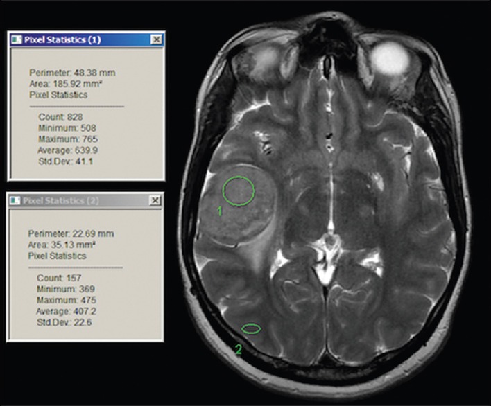
Materials and methods: A prospective study included all the patients who were admitted for surgery with an MRI finding suggestive of meningioma. All patients were subjected to routine radiological investigations. Surgical resection was performed for patients eligible for surgery using cavitron ultrasonic aspirator (CUSA). The relationship and correlation between the radiological, intraoperative measurements and the histopathological diagnosis were studied. The tumor consistency was measured using mean CUSA level. Intensity on T2, fluid-attenuated inversion recovery (FLAIR), and diffusion-weighted imaging (DWI) was measured using circular regions of interest (ROI) on the MRI. Multiple ROIs were placed initially on the lesions avoiding the obvious blood vessels, if any, then on the brain cortex to avoid the vasogenic edema. The mean ROI (mROI) results from the lesion were subtracted from the mean ROI from the brain cortex for each lesion to achieve normalized ratio. The results of lesion mROI-cortex mROI were compared to the operative and histopathology results using Pearson's correlation test and linear regression test.
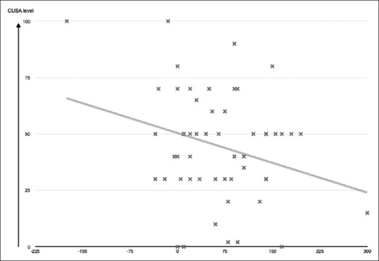
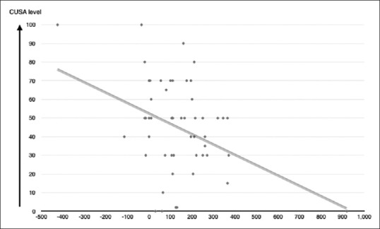
Results: The total number of patients was seventy. The mean age of the patients was 51 ± 14.8, with 72% of them being females and 28% males. There was a strong statistically significant (P = 0.046) and (P = 0.003) correlation between mean CUSA and FLAIR mROI difference or T2 mROI difference, respectively. On the other hand, there was an inversely proportional relationship between mean CUSA and FLAIR mROI difference and mean CUSA and T2 mROI difference. The value of the regression test (r) shows that there was a slight linear relationship between FLAIR mROI difference or T2 mROI difference and mean CUSA values, in which the mean CUSA value = 50.1 + (-0.088) × FLAIR mROI difference (r = -0.273, P = 0.046) or mean CUSA value = 50.8 + (-0.055) × T2 mROI difference (r = 0.4, P = 0.003). There was no statistical significance in the relation between CUSA values and tumor histological subtypes, DWI values, age, or gender.
Conclusion: This study presents a new objective method to measure the consistency of intracranial meningiomas based on a simple algorithmic formula. Such information will aid in planning surgery and assessing the resectability of the tumor. To date, this is the first objective measurement of meningioma consistency based on MRI studies and objective intraoperative evaluation.
Keywords: Brain imaging; magnetic resonance imaging; meningioma; neurostimulation; neurosurgery.
Conflict of interest statement
There are no conflicts of interest.
Figures
Similar articles
-
Meningioma consistency prediction utilizing tumor to cerebellar peduncle intensity on T2-weighted magnetic resonance imaging sequences: TCTI ratio.J Neurosurg. 2017 Jan;126(1):242-248. doi: 10.3171/2016.1.JNS152329. Epub 2016 Apr 8. J Neurosurg. 2017. PMID: 27058200
-
The Predictive Value of Conventional Magnetic Resonance Imaging Sequences on Operative Findings and Histopathology of Intracranial Meningiomas: A Prospective Study.Neurol India. 2019 Nov-Dec;67(6):1439-1445. doi: 10.4103/0028-3886.273632. Neurol India. 2019. PMID: 31857531
-
Supratentorial Meningioma Consistency Prediction Utilizing Tumor to Cerebellar Peduncle Intensity on T1 and T2-Weighted and Fluid Attenuated Inversion Recovery Magnetic Resonance Imaging Sequences.World Neurosurg. 2023 Feb;170:e180-e187. doi: 10.1016/j.wneu.2022.10.097. Epub 2022 Nov 1. World Neurosurg. 2023. PMID: 36328167
-
Benefits of Combined MRI Sequences in Meningioma Consistency Prediction: A Prospective Study of 287 Consecutive Patients.Asian J Neurosurg. 2022 Dec 10;17(4):614-620. doi: 10.1055/s-0042-1758849. eCollection 2022 Dec. Asian J Neurosurg. 2022. PMID: 36570751 Free PMC article.
-
Intracranial meningiomas: correlations between MR imaging and histology.Eur J Radiol. 1999 Jul;31(1):69-75. doi: 10.1016/s0720-048x(98)00083-7. Eur J Radiol. 1999. PMID: 10477102 Review.
Cited by
-
Differentiation between various types and subtypes of intracranial meningiomas with advanced MRI.SA J Radiol. 2022 Oct 26;26(1):2480. doi: 10.4102/sajr.v26i1.2480. eCollection 2022. SA J Radiol. 2022. PMID: 36337074 Free PMC article.
-
Predicting intraoperative meningioma consistency using features from standard MRI sequences: a preoperative evaluation.Acta Neurochir (Wien). 2025 Jun 21;167(1):173. doi: 10.1007/s00701-025-06582-9. Acta Neurochir (Wien). 2025. PMID: 40542946 Free PMC article.
-
Utility of texture analysis for objective quantitative ex vivo assessment of meningioma consistency: method proposal and validation.Acta Neurochir (Wien). 2023 Dec;165(12):4203-4211. doi: 10.1007/s00701-023-05867-1. Epub 2023 Dec 4. Acta Neurochir (Wien). 2023. PMID: 38044374
-
Histogram analysis of mono-exponential, bi-exponential and stretched-exponential diffusion-weighted MR imaging in predicting consistency of meningiomas.Cancer Imaging. 2023 Dec 5;23(1):117. doi: 10.1186/s40644-023-00633-z. Cancer Imaging. 2023. PMID: 38053183 Free PMC article.
-
Isolated incomplete third cranial nerve palsy due to presumed cavernous sinus meningioma.Oman J Ophthalmol. 2022 Mar 2;15(1):104-106. doi: 10.4103/ojo.ojo_406_20. eCollection 2022 Jan-Apr. Oman J Ophthalmol. 2022. PMID: 35388256 Free PMC article. No abstract available.
References
-
- Claus EB, Bondy ML, Schildkraut JM, Wiemels JL, Wrensch M, Black PM. Epidemiology of intracranial meningioma. Neurosurgery. 2005;57:1088–95. - PubMed
-
- Jääskeläinen J, Haltia M, Servo A. Atypical and anaplastic meningiomas: Radiology, surgery, radiotherapy, and outcome. Surg Neurol. 1986;25:233–42. - PubMed
-
- Central Brain Tumor Registry of the United States. Statistical report: Primary brain tumors in the United States, 1998-2002. Hinsdale, Illinois: Central Brain Tumor Registry of the United States; 2005.
LinkOut - more resources
Full Text Sources
Other Literature Sources