. 2017 Jun;7(2):1750005.
doi: 10.1142/s1793984417500052.

Prostaglandin E2 Produced by Alginate-Encapsulated Mesenchymal Stromal Cells Modulates the Astrocyte Inflammatory Response

Affiliations

Prostaglandin E2 Produced by Alginate-Encapsulated Mesenchymal Stromal Cells Modulates the Astrocyte Inflammatory Response

Elizabeth C Stucky et al. Nano Life. 2017 Jun.

Abstract

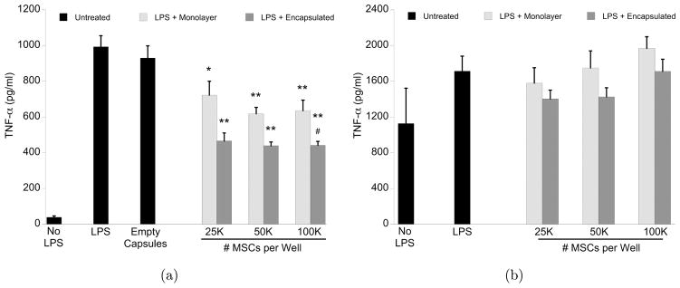
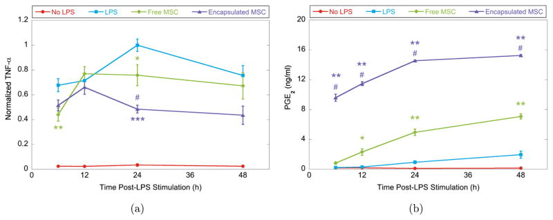
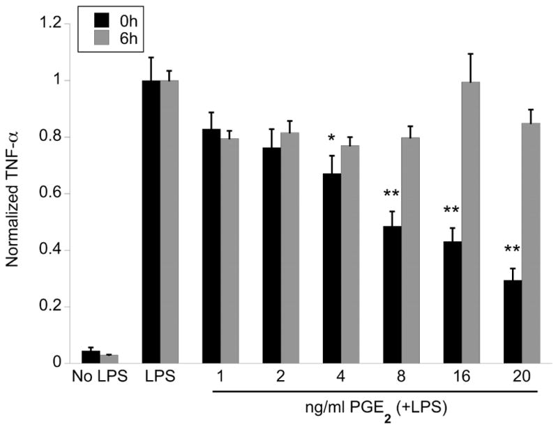
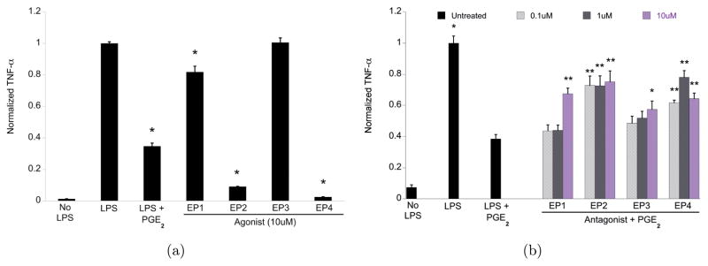
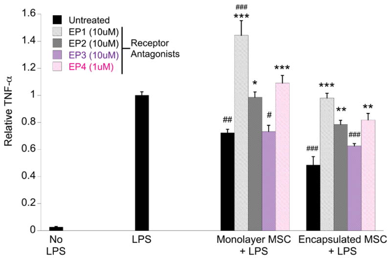
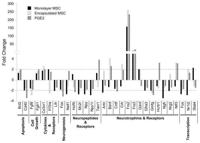
Astroglia are well known for their role in propagating secondary injury following brain trauma. Modulation of this injury cascade, including inflammation, is essential to repair and recovery. Mesenchymal stromal cells (MSCs) have been demonstrated as trophic mediators in several models of secondary CNS injury, however, there has been varied success with the use of direct implantation due to a failure to persist at the injury site. To achieve sustained therapeutic benefit, we have encapsulated MSCs in alginate microspheres and evaluated the ability of these encapsulated MSCs to attenuate neuro-inflammation. In this study, astroglial cultures were administered lipopolysaccharide (LPS) to induce inflammation and immediately co-cultured with encapsulated or monolayer human MSCs. Cultures were assayed for the pro-inflammatory cytokine tumor necrosis factor alpha (TNF-α) produced by astroglia, MSC-produced prostaglandin E2, and expression of neurotrophin-associated genes. We found that encapsulated MSCs significantly reduced TNF-α produced by LPS-stimulated astrocytes, more effectively than monolayer MSCs, and this enhanced benefit commences earlier than that of monolayer MSCs. Furthermore, in support of previous findings, encapsulated MSCs constitutively produced high levels of PGE2, while monolayer MSCs required the presence of inflammatory stimuli to induce PGE2 production. The early, constitutive presence of PGE2 significantly reduced astrocyte-produced TNF-α, while delayed administration had no effect. Finally, MSC-produced PGE2 was not only capable of modulating inflammation, but appears to have an additional role in stimulating astrocyte neurotrophin production. Overall, these results support the enhanced benefit of encapsulated MSC treatment, both in modulating the inflammatory response and providing neuroprotection.

Keywords: Astroglia; inflammatory mediators; mesenchymal stromal cells; prostaglandin E2; traumatic brain injury.


Figures

Fig. 1
Fig. 1
Rat TNF-α ELISA of cell culture media supernatant collected after 24 h of LPS stimulation ±MSC treatment in astrocyte (a) and microglia (b) cultures. Data are represented as mean ±S.E. from three experiments, each with N = 2–3 cultures per condition. In astrocyte culture, encapsulated MSC treatment significantly reduced TNF-α levels, and was more effective than monolayer MSC treatment at the highest dose evaluated. Empty capsule treatment had no significant effect on TNF-α reduction. MSC treatment had no effect in microglia cultures. *= p < 0.02, **= p < 0.002, ***= p < 0.0001 compared to LPS + no treatment, #= p < 0.01, ##= p < 0.002 compared to treatment with equivalent number of monolayer MSC.
Fig. 2
Fig. 2
Temporal profile of rat TNF-α and total PGE2 levels in culture media collected after LPS stimulation ± MSC treatment in astrocytes. TNF-α data are normalized to untreated LPS-stimulated cultures at 24 h. All data are represented as mean ± S.E. from three experiments, each with N = 3 cultures per condition. (a) Encapsulated MSC treatment shows an early trend in reducing TNF-α more effectively than monolayer MSCs, which is maintained to the 48 h endpoint. *= p < 0.05, **= p < 0.01, ***= p < 0.0001 compared to LPS only, #= p < 0.05 compared to LPS + monolayer MSC. (b) High levels of PGE2 are produced by encapsulated MSCs from 6 h post-stimulation, whereas monolayer MSCs start producing PGE2 at significantly lower levels from 12 h post-stimulation. *= p < 0.001, **= p < 0.0001 compared to LPS only, #= p < 0.0001 compared to LPS + monolayer MSC.
Fig. 3
Fig. 3
Rat TNF-α ELISA of cell culture media collected from astrocyte cultures after 24 h of LPS stimulation ±human PGE2. Data is normalized to untreated LPS-stimulated astrocytes and represented as mean ± S.E. from three experiments, each with N = 3 cultures per condition. Addition of exogenous human PGE2 significantly reduced TNF-α levels in a dose dependent manner when immediately administered, but had no effect when administered 6 h after LPS. *= p < 0.01, **= p < 0.0001 compared to LPS only.
Fig. 4
Fig. 4
Effect of PGE2 receptor subtype-specific agonists and antagonists on TNF-α reduction. Data are normalized to untreated LPS-stimulated astrocytes and represented as mean ± S.E. from three experiments, each with N = 3 cultures per condition. (a) Rat TNF-α produced by astrocyte cultures after 24 h of LPS stimulation ± EP receptor agonist iloprost (EP1), butaprost (EP2), sulprostone (EP3), or CAY1058 (EP4). A significant, strong agonist effect is observed for the EP2 and EP4 receptors, and a milder, but significant effect for the EP1 receptor. No effect is seen on the EP3 receptor. *= p < 0.0001 compared to LPS only. (b) Rat TNF-α produced by astrocytes after 24 h of LPS stimulation + 20 ng/ml PGE2 ± EP receptor antagonist SC-51322 (EP1), PF-04418948 (EP2), L-798,106 (EP3), or L-161,982 (EP4). Significant antagonist blocking is observed for all EP receptor subtypes. *= p < 0.05, **= p < 0.0005 compared to LPS + PGE2.
Fig. 5
Fig. 5
PGE2 receptor antagonist blocking of MSC treatment. Rat TNF-α produced by astrocytes after 24 h of LPS stimulation + MSC (monolayer or encapsulated) ± EP receptor antagonist SC-51322 (EP1), PF-04418948 (EP2), L-798,106 (EP3), or L-161,982 (EP4). Significant blocking of MSC-mediated TNF-α reduction was observed with antagonists specific for the EP1, EP2, and EP4 receptors. No effect was seen using the EP3 receptor-specific antagonist. *= p < 0.05, **= p < 0.005, ***= p < 0.0005 compared to MSC only counterpart, #= p < 0.05, ##= p < 0.005, ###= p < 0.0001 compared to LPS only.
Fig. 6
Fig. 6
Fold changes in astrocyte neurotrophin-associated gene expression after MSC or PGE2 treatment, for 30 genes (of 84 assayed) that exhibited at least two-fold up- or down-regulation (dashed line) in one or more treatment conditions evaluated, relative to untreated, LPS-stimulated astrocytes.

Similar articles

Cited by

References

    1. Markiewicz I, Lukomska B. Acta Neurobiol Exp. 2006;66:343. - PubMed
    1. Nakajima K, Kohsaka S. J Biochem. 2001;130:169. - PubMed
    1. Benveniste EN. Am J Physiol. 1992;263:C1. - PubMed
    1. Shastri A, Bonifati DM, Kishore U. Mediators Inflamm. 2013;2013:1. - PMC - PubMed
    1. Zhukareva V, Obrocka M, Houle JD, Fischer I, Neuhuber B. Cytokine. 2010;50:317. - PubMed

LinkOut - more resources