

Primary External Iliac Venous Aneurysm: A Case Report
- PMID: 29682124
- PMCID: PMC5882340
- DOI: 10.3400/avd.cr.17-00080
Primary External Iliac Venous Aneurysm: A Case Report
Abstract
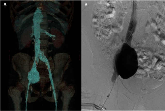
Primary iliac venous aneurysm is an exceedingly rare abnormality that can be complicated by pulmonary embolism, thrombosis, and rupture. Here we report the case of an otherwise healthy 40-year-old man with a unilateral external iliac vein aneurysm without any evidence of an arteriovenous fistula, proximal stenosis, or obstruction, as reported on computed tomography. Pulmonary embolism was diagnosed using 99mTc-macroaggregated albumin scintigraphy. To prevent life-threatening complications, we treated the patient with anticoagulant therapy and performed aneurysmectomy with reconstruction using a saphenous vein graft patch. Although postoperative venography showed obstruction of the external iliac vein, the patient remained asymptomatic.
Keywords: iliac vein; primary venous aneurysm; treatment.
Figures

Similar articles
-
Ruptured aneurysm of the external iliac vein.J Vasc Surg Venous Lymphat Disord. 2016 Jan;4(1):92-4. doi: 10.1016/j.jvsv.2015.07.005. J Vasc Surg Venous Lymphat Disord. 2016. PMID: 26946902
-
Pulmonary embolism caused by a thrombosed external iliac venous aneurysm.Ann Vasc Surg. 2011 Oct;25(7):982.e15-8. doi: 10.1016/j.avsg.2011.03.013. Epub 2011 Jun 15. Ann Vasc Surg. 2011. PMID: 21680142
-
External iliac venous aneurysm in a pregnant woman: a case report.J Vasc Surg. 2004 Jul;40(1):174-8. doi: 10.1016/j.jvs.2004.02.043. J Vasc Surg. 2004. PMID: 15218481 Review.
-
Simultaneous Iliac Vein Bovine Pericardial Patch Venoplasty and Creation of PTFE Lower Limb Arteriovenous Fistula Graft for Rescue Vascular Access.Ann Vasc Surg. 2016 Oct;36:292.e9-292.e11. doi: 10.1016/j.avsg.2016.03.018. Epub 2016 Jul 15. Ann Vasc Surg. 2016. PMID: 27423716
-
[Iliac venous aneurysm: a case report and review of literature].Chirurgia (Bucur). 2011 Mar-Apr;106(2):269-72. Chirurgia (Bucur). 2011. PMID: 21698869 Review. Romanian.
Cited by
-
Open aneurysmorrhaphy for repair of a massive iliac vein aneurysm and review of the recent literature.J Vasc Surg Cases Innov Tech. 2023 Sep 28;9(4):101336. doi: 10.1016/j.jvscit.2023.101336. eCollection 2023 Dec. J Vasc Surg Cases Innov Tech. 2023. PMID: 37885794 Free PMC article.
-
Giant renal vein aneurysm.J Vasc Surg Cases Innov Tech. 2019 Aug 7;5(3):365-368. doi: 10.1016/j.jvscit.2019.06.007. eCollection 2019 Sep. J Vasc Surg Cases Innov Tech. 2019. PMID: 31440715 Free PMC article.
-
[Management of venous aneurysms and the vascular surgical treatment options : Selection of representative case constellations illustrating experiences at a center for vascular surgery].Chirurgie (Heidelb). 2025 Jul;96(7):583-592. doi: 10.1007/s00104-024-02191-x. Epub 2024 Nov 14. Chirurgie (Heidelb). 2025. PMID: 39542913 Free PMC article. German.
-
Phlegmasia cerulea dolens secondary to an aortoiliac aneurysm.J Vasc Surg Cases Innov Tech. 2019 Jun 27;5(3):278-282. doi: 10.1016/j.jvscit.2019.03.008. eCollection 2019 Sep. J Vasc Surg Cases Innov Tech. 2019. PMID: 31312778 Free PMC article.
References
-
- Ysa A, Bustabad MR, Arruabarrena A, et al. Thrombosed iliac venous aneurysm: a rare form of presentation of a congenital anomaly of the inferior vena cava. J Vasc Surg 2008; 48: 218-22. - PubMed
-
- Postma MP, McLellan GL, Northup HM, et al. Aneurysm of the internal iliac vein as a rare source of pulmonary thromboembolism. South Med J 1989; 82: 390-2. - PubMed
-
- Petrunić M, Kruzić Z, Tonković I, et al. Large iliac venous aneurysm simulating a retroperitoneal soft tissue tumour. Eur J Vasc Endovasc Surg 1997; 13: 221-2. - PubMed
-
- Alatri A, Radicchia S. Bilateral aneurysm of the common iliac vein: a case report. Ann Ital Med Int 1997; 12: 92-3. (in Italian) - PubMed
-
- Fourneau I, Reynders-Frederix V, Lacroix H, et al. Aneurysm of the iliofemoral vein. Ann Vasc Surg 1998; 12: 605-8. - PubMed
Publication types
LinkOut - more resources
Full Text Sources
Other Literature Sources