Mandible protraction alters Type I collagen, osteocalcin and osteonectin gene expression in adult mice condyle
- PMID: 29682221
- PMCID: PMC5897095
- DOI: 10.11138/ads/2017.8.3.095
Mandible protraction alters Type I collagen, osteocalcin and osteonectin gene expression in adult mice condyle
Abstract
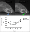
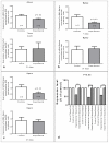
Mandible condyle remodeling is a great challenge on craniofacial growth studies. The great majority of the reports deals with growing period. However, there is a great necessity to clarify the importance of functional stimulation on adult mandible condyle remodeling. By using an adult mouse model, we investigated the influence of mandible forwarding on condyle remodeling and gene expression by bone forming cells. Tomographic and scintigraphic evaluations showed sagittal growth and cell activity enhancement. RT-PCR showed that Type I collagen, osteocalcin and osteonectin expression level can be altered. We showed that functional stimulation is necessary to maintain the regular gene expression by condyle bone forming cells in adult mice. It opens new frame for further investigations aiming new clinical approaches to temporomandibular joint problems treatment, as well as mandible retrusion treatment.
Keywords: CT; bone biology; condyle growth; gene expression; growth evaluation; molecular biology.
Figures


Similar articles
-
Effects of transmandibular symphyseal distraction on teeth, bone, and temporomandibular joint.J Oral Maxillofac Surg. 2009 Oct;67(10):2254-65. doi: 10.1016/j.joms.2009.04.055. J Oral Maxillofac Surg. 2009. PMID: 19761921
-
[The preliminary study on the characteristics of mandible and condyle in dentin matrix protein-1 gene knockout mice].Zhonghua Kou Qiang Yi Xue Za Zhi. 2005 Jul;40(4):335-7. Zhonghua Kou Qiang Yi Xue Za Zhi. 2005. PMID: 16191382 Chinese.
-
[Use of condyle positioning plate in sagittal split ramus osteotomy of mandible].Zhonghua Kou Qiang Yi Xue Za Zhi. 1996 May;31(3):165-8. Zhonghua Kou Qiang Yi Xue Za Zhi. 1996. PMID: 9387560 Chinese.
-
The stock alloplastic temporomandibular joint implant can influence the behavior of the opposite native joint: A numerical study.J Craniomaxillofac Surg. 2015 Oct;43(8):1384-91. doi: 10.1016/j.jcms.2015.06.042. Epub 2015 Jul 8. J Craniomaxillofac Surg. 2015. PMID: 26231883
-
Temporomandibular Joint Anatomy Assessed by CBCT Images.Biomed Res Int. 2017;2017:2916953. doi: 10.1155/2017/2916953. Epub 2017 Feb 2. Biomed Res Int. 2017. PMID: 28261607 Free PMC article. Review.
Cited by
-
Evaluation of the Salivary Expression of Type I Collagen, Osteocalcin, and Osteonectin in Patients Treated With Myofunctional Therapy: A Clinical Study.Cureus. 2025 Aug 10;17(8):e89762. doi: 10.7759/cureus.89762. eCollection 2025 Aug. Cureus. 2025. PMID: 40799669 Free PMC article.
References
LinkOut - more resources
Full Text Sources
Other Literature Sources
