Submandibular lipoblastoma: Case report of a rare tumor in childhood
- PMID: 29682479
- PMCID: PMC5898184
- DOI: 10.4103/ajm.AJM_81_17
Submandibular lipoblastoma: Case report of a rare tumor in childhood
Abstract
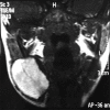
Lipoblastoma is a rare, benign tumor usually occurring in childhood. It is essentially localized in the extremities and trunk, with few cases reported in the neck. We report the case of a 2-year-old girl with a rapidly enlarging, painless neck mass. Magnetic resonance imaging (MRI) revealed a 3-cm mass in the right submandibular region. Review of literature, diagnostic methods, and genetics of lipomatous tumors are discussed. Complete surgical excision via a lateral cervical approach demonstrated a white soft tissue with an adherent ganglion. Histology and immunohistochemistry confirmed the diagnosis of lipoblastoma. Cervical lipoblastoma is rare, and typically asymptomatic, rarely causing nerve compression or airway obstruction. MRI can help identifying the lipomatous nature of the mass, but the findings can be inconsistent due to variable maturity of fat cells and the mesenchymal content of the tumor. Diagnosis is always based on pathological examination. Further chromosomal analysis is useful in differentiating lipoblastoma from liposarcoma. Complete surgical excision is the recommended treatment.
Keywords: Benign tumor; head and neck; immunohistochemestry; lipoblastoma; liposarcoma.
Conflict of interest statement
There are no conflicts of interest.
Figures


Similar articles
-
Cervical lipoblastoma: case report, review of literature, and genetic analysis.Head Neck. 2007 Nov;29(11):1055-60. doi: 10.1002/hed.20633. Head Neck. 2007. PMID: 17427967 Review.
-
Head and neck lipoblastomas: Report of 3 cases and review of the literature.Int J Surg Case Rep. 2021 Jul;84:106050. doi: 10.1016/j.ijscr.2021.106050. Epub 2021 Jun 4. Int J Surg Case Rep. 2021. PMID: 34139421 Free PMC article.
-
Pediatric lipoblastoma of the neck.J Craniofac Surg. 2013;24(5):e507-10. doi: 10.1097/scs.0b013e31828dcf71. J Craniofac Surg. 2013. PMID: 24163863 Review.
-
Perineal lipoblastoma: a case report and review of literature.Int J Clin Exp Pathol. 2014 May 15;7(6):3370-4. eCollection 2014. Int J Clin Exp Pathol. 2014. PMID: 25031762 Free PMC article. Review.
-
Shoulder Lipoblastoma in a 2-Year-Old Boy Case Report and Literature Review.J Orthop Case Rep. 2021 Dec;11(12):84-87. doi: 10.13107/jocr.2021.v11.i12.2580. J Orthop Case Rep. 2021. PMID: 35415147 Free PMC article.
Cited by
-
Rapidly Growing Facial Tumor in a 5-Year-Old Girl.Ann Maxillofac Surg. 2020 Jan-Jun;10(1):267-271. doi: 10.4103/ams.ams_200_19. Epub 2020 Jun 8. Ann Maxillofac Surg. 2020. PMID: 32855956 Free PMC article.
-
Imaging of head and neck lipoblastoma: case report and systematic review.J Ultrasound. 2021 Sep;24(3):231-239. doi: 10.1007/s40477-020-00439-w. Epub 2020 Mar 5. J Ultrasound. 2021. PMID: 32141045 Free PMC article.
References
-
- Chung EB, Enzinger FM. Benign lipoblastomatosis. An analysis of 35 cases. Cancer. 1973;32:482–92. - PubMed
-
- Basaran UN, Inan M, Bilgi S, Pul M. Lipoblastoma: A rare cervical mass in childhood. Int J Pediatr Otorhinolaryngol. 2001;61:265–8. - PubMed
-
- Rasmussen IS, Kirkegaard J, Kaasbøl M. Intermittent airway obstruction in a child caused by a cervical lipoblastoma. Acta Anaesthesiol Scand. 1997;41:945–6. - PubMed
-
- O'Donnell KA, Caty MG, Allen JE, Fisher JE. Lipoblastoma: Better termed infantile lipoma? Pediatr Surg Int. 2000;16:458–61. - PubMed
-
- Farrugia MK, Fearne C. Benign lipoblastoma arising in the neck. Pediatr Surg Int. 1998;13:213–4. - PubMed
Publication types
LinkOut - more resources
Full Text Sources
Other Literature Sources

