. 2018 Feb 28:2018:9508721.
doi: 10.1155/2018/9508721. eCollection 2018.

Antibody-Mediated Osseous Regeneration for Bone Tissue Engineering in Canine Segmental Defects

Affiliations

Antibody-Mediated Osseous Regeneration for Bone Tissue Engineering in Canine Segmental Defects

A Khojasteh et al. Biomed Res Int. .

Abstract

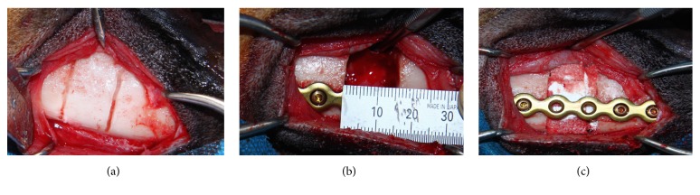
Among many applications of therapeutic monoclonal antibodies (mAbs), a unique approach for regenerative medicine has entailed antibody-mediated osseous regeneration (AMOR). In an effort to identify a clinically relevant model of craniofacial defect, the present study investigated the efficacy of mAb specific for bone morphogenetic protein- (BMP-) 2 to repair canine segmental mandibular continuity defect model. Accordingly, a 15 mm unilateral segmental defect was created in mandible and fixated with a titanium plate. Anorganic bovine bone mineral with 10% collagen (ABBM-C) was functionalized with 25 μg/mL of either chimeric anti-BMP-2 mAb or isotype-matched mAb (negative control). Recombinant human (rh) BMP-2 served as positive control. Morphometric analyses were performed on computed tomography (CT) and histologic images. Bone densities within healed defect sites at 12 weeks after surgery were 1360.81 ± 10.52 Hounsfield Unit (HU), 1044.27 ± 141.16 HU, and 839.45 ± 179.41 HU, in sites with implanted anti-BMP-2 mAb, rhBMP-2, and isotype mAb groups, respectively. Osteoid bone formation in anti-BMP-2 mAb (42.99% ± 8.67) and rhBMP-2 (48.97% ± 2.96) groups was not significantly different but was higher (p < 0.05) than in sites with isotype control mAb (26.8% ± 5.35). In view of the long-term objective of translational application of AMOR in humans, the results of the present study demonstrated the feasibility of AMOR in a large clinically relevant animal model.


Figures

Figure 1
Figure 1
Intraoperative clinical images of the steps in segmental osteotomy and repair. Extraoral surgical approach to expose the mandible and depth grooving to mark the exact defect size (a). Fixation of mandibular segments with titanium reconstruction plate (b). Segmental mandibulectomy (15 mm) was performed and a titanium plate was used to rigidly fixate the two segments together. The resected mandibular segmental defect was implanted with anorganic bovine bone mineral collagen, functionalized with anti-BMP-2 mAb or isotype-matched control mAb (c).
Figure 2
Figure 2
CT scan images of the bone formation within canine segmental defects. Three-dimensional images of the defects after 6 weeks in isotype mAb (a1), rhBMP-2 (b1), and anti-BMP-2 (c1) groups. Coronal sections of the defect's center slide were shown at 6th week in isotype mAb (a2), rhBMP-2 (b2), and anti-BMP-2 mAb (c2) groups. Same sections at 12th week in isotype mAb (a3), rhBMP-2 (b3), and anti-BMP-2 (c3) groups.
Figure 3
Figure 3
Quantitative analysis of calcified tissues in the defect sites by CT scan imaging. (a) Comparison of bone density (Hounsfield Unit) of isotype mAb, rhBMP-2, and anti-BMP-2 mAb at 6th and 12th weeks. (b) Comparison of mineralized area (mm2) of isotype mAb, rhBMP-2, and anti-BMP-2 mAb at 6th and 12th weeks. Means and standard deviations were calculated in each group and statistical significance was assessed by general linear models for repeated measures and Tukey's test as the post hoc test (,•,O,†p < 0.05; all other comparisons in each time point were not significant).
Figure 4
Figure 4
Histological examination of specimens at 12 weeks postoperatively. (a) Schematic diagram of the segmental defects, illustrating the location of sections taken for histological analysis. The proximal and distal segments were sectioned horizontally and the cross section of the central segment was taken (b, f, j). (b–m) Representative histomicrograms stained with H&E (40x). (b–e) Isotype-matched mAb, (f–i) rhBMP-2, and (j–m) anti-BMP-2 mAb used to functionalize ABBM-C scaffold. The blue arrows show the junction of newly formed bone and the neighboring host bone. The white arrows show the residual scaffold biomaterials, which were surrounded by connective tissue in control specimens treated with isotype-matched control mAb, while in experimental and positive control specimens the residual scaffold was not easily discernable. Abundant endothelial-lined blood vessels (green arrows) were noted in anti-BMP-2 mAb and rhBMP-2 treated sites.
Figure 5
Figure 5
Quantitative histomorphometric analysis of bone specimens for bone volume/total volume (%) and residual graft (%). Means and standard deviations of each group were calculated and statistical significance was assessed by ANOVA and Tukey's post hoc test.

References

    1. Martino M. M., Briquez P. S., Maruyama K., Hubbell J. A. Extracellular matrix-inspired growth factor delivery systems for bone regeneration. Advanced Drug Delivery Reviews. 2015;94:41–52. doi: 10.1016/j.addr.2015.04.007. - DOI - PubMed
    1. Behnia H., Khojasteh A., Soleimani M., Tehranchi A., Atashi A. Repair of alveolar cleft defect with mesenchymal stem cells and platelet derived growth factors: a preliminary report. Journal of Cranio-Maxillo-Facial Surgery. 2012;40(1):2–7. doi: 10.1016/j.jcms.2011.02.003. - DOI - PubMed
    1. Khojasteh A., Eslaminejad M. B., Nazarian H. Mesenchymal stem cells enhance bone regeneration in rat calvarial critical size defects more than platelete-rich plasma. Oral Surgery, Oral Medicine, Oral Pathology, Oral Radiology, and Endodontology. 2008;106(3):356–362. doi: 10.1016/j.tripleo.2007.10.017. - DOI - PubMed
    1. Kempen D. H. R., Creemers L. B., Alblas J., et al. Growth Factor interactions in bone regeneration. Tissue Engineering - Part B: Reviews. 2010;16(6):551–566. doi: 10.1089/ten.teb.2010.0176. - DOI - PubMed
    1. Stevenson S., Cunningham N., Toth J., Davy D., Reddi A. H. The effect of osteogenin (a bone morphogenetic protein) on the formation of bone in orthotopic segmental defects in rats. The Journal of Bone & Joint Surgery. 1994;76(11):1676–1687. doi: 10.2106/00004623-199411000-00011. - DOI - PubMed

LinkOut - more resources