. 2018 Apr 9;4(4):1241-1250.
doi: 10.1021/acsbiomaterials.6b00706. Epub 2017 Feb 20.

Dual Affinity Heparin-Based Hydrogels Achieve Pro-Regenerative Immunomodulation and Microvascular Remodeling

Affiliations

Dual Affinity Heparin-Based Hydrogels Achieve Pro-Regenerative Immunomodulation and Microvascular Remodeling

Molly E Ogle et al. ACS Biomater Sci Eng. .

Abstract

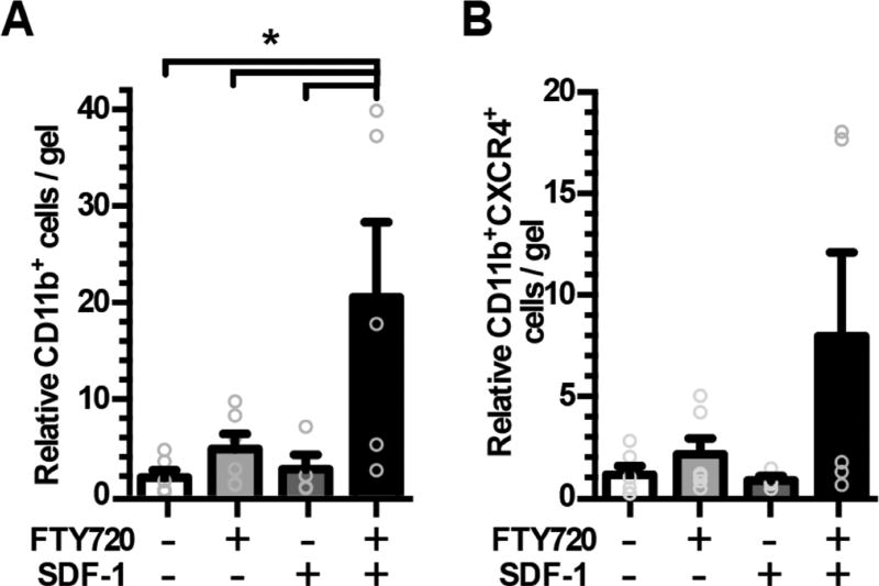
The immune response to biomaterial implants critically regulates functional outcomes such as vascularization, transplant integration/survival, and fibrosis. To create "immunologically smart" materials, the host-material response may be engineered to optimize the recruitment of pro-regenerative leukocyte subsets which mature into corresponding wound-healing macrophages. We have recently identified a unique feature of pro-regenerative Ly6Clow monocytes that is a higher expression of both the bioactive lipid receptor sphingosine-1-phosphate receptor 3 (S1PR3) and the stromal derived factor-1α (SDF-1α) receptor CXCR4. Therefore, we designed a bifunctional hydrogel to harnesses a mechanistic synergy between these signaling axes to enhance the recruitment of endogenous pro-regenerative monocytes. To overcome the challenge of codelivering two physiochemically distinct molecules-a large hydrophilic protein and hydrophobic small molecule-we engineered a dual affinity hydrogel that exploits the growth factor affinity of a heparin derivative (Hep-N) and lipid chaperone activity of albumin. The sphingosine analog FTY720 and SDF-1α are successfully loaded and coreleased from the Hep-N-functionalized PEG-DA hydrogels while maintaining bioactivity. Placement of these hydrogels into a murine partial thickness skin wound demonstrates that corelease of FTY720 and SDF-1α yields superior recruitment of myeloid cells to the implant interface compared to either factor alone. Although in vivo delivery of FTY720 or SDF-1α individually promotes the enhanced recruitment of Ly-6Clow anti-inflammatory monocytes, codelivery enhances the early accumulation and persistence of the differentiated wound healing CD206+ macrophages in the tissue surrounding the gel. Co-delivery similarly promoted the synergistic expansion of vasculature adjacent to the implant, a key step in tissue healing. Taken together, these findings suggest that the combination of chemotactic molecules may provide additional maturation signals to the infiltrating leukocytes to facilitate macrophage transition and vascular network expansion, thus, ultimately, potentiating tissue repair. The coupling of multiple pro-regenerative biological cues provides a foundation for more fine-tuned immunoregenerative modulation to facilitate tissue repair.

Keywords: bioactive lipids; heparin hydrogels; immunoregenerative engineering; stromal derived factor-1α (SDF-1α).


Conflict of interest statement

The authors declare no competing financial interest.

Figures

Figure 1
Figure 1
Hep−N-PEG-DA hydrogels corelease bioactive SDF-1α and FTY720 in vitro. (A) Fabrication and postgelation loading of SDF-1α and/or FTY720 into albumin-embedded Hep−N-PEG-DA gels (aHep−N). (B) SDF-1α and FTY720 are released over 7 days in vitro (n = 6 gels, 2 independent studies). (C) In vitro migration of primary murine bone marrow cells toward media conditioned by 24h of hydrogel release. *Indicates significance compared to all other groups (n = 5, ANOVA, p < 0.05).
Figure 2
Figure 2
Dual release of SDF-1α and FTY720 from aHep−N-PEG-DA hydrogels promotes synergistic recruitment of leukocytes to the gel surface. Gels were implanted in the dorsal skin window chamber for 3 days and explanted to evaluate cell association with the gel by flow cytometric analysis of (A) CD11b+ and (B) CD11b+CXCR4+ populations. (n = 5–6, ANOVA, *p < 0.05).
Figure 3
Figure 3
Myeloid cells have greater directional motility around dual releasing gel. Myeloid cell movements were captured by intra vital microscopy of the dorsal window chamber in the CX3CR1GFP/+ transgenic mouse model 1 day after surgery and gel implantation. Myeloid cells (green) and vessels (red) were visible adjacent to the unloaded Hep−N-PEG-DA internal control gel (A) and the dual releasing aHep−N-PEG-DA SDF-1α + FTY720 gel (B) in the same window chamber. Fifteen minute videos were acquired and cell tracking analysis was done in IMARIS software. Cell displacement (C) was enhanced in the region surrounding the experimental gel but not track length (D), suggesting that the cells are traveling in a straighter and more directed path (E). (n = 2119–3095 cells, 2 mice/group, 2 regions of interest each, Mann-Whitney rank test, *p < 0.05).
Figure 4
Figure 4
Recruitment of mononuclear phagocyte subsets to tissue surrounding aHep−N-PEG-DA gels. Tissue surrounding the hydrogel was analyzed 3 days after surgery and implantation for the immunophenotype of the recruited myeloid populations. (A) Either FTY720 or SDF-1α lead to an increase in AM (SSClowCD11b+Ly6Clow) recruitment to the gel as a percent of monocytes (SSClowCD11b+) (n = 7–9, ANOVA, *p < 0.05). (B) Dual release of FTY720 and SDF-1α significantly increased the presence of MerTK+CD64+ macrophages within the tissue as a percent of total cells (n = 2–4, ANOVA of control, SDF-1, and FTY720/SDF-1 groups (FTY720 excluded due to low sample size)). (C) Dual release similarly produced a trend of increased CD206+MerTK+CD64+ cells as a percent of total cells (n = 2–4, ANOVA of control, SDF-1, and FTY720/SDF-1 groups, p = 0.07).
Figure 5
Figure 5
Dual release of SDF-1α and FTY720 alters CX3CR1+ cell morphology. (A) CX3CR1−GFP/+ mice were fitted with a dorsal window chamber and after 6 days were injected (i.v.) with rhodamine-dextran to visualize perfused vessels. Confocal intravital imaging was then conducted in the region directly surrounding the hydrogel implant. (B, D) Tissue surrounding the SDF hydrogel had rounded CX3CR1-GFP+ cells surrounding the vasculature, whereas (C, E) tissue surrounding the SDF-1α + FTY720 hydrogel had CX3CR1dim myeloid cells with elongated morphology consistent with macrophage phenotype. Scale bar 100 µm. (representative images from n = 3 ROI per gel).
Figure 6
Figure 6
Dual release of SDF-1 and FTY720 increases abundance of CD206+ macrophages. (A) Whole-mount IHC and confocal microscopy shows accumulation of CD68+ macrophages and CD206+CD68+ macrophages in tissue proximal to the gels at day 7 (CD206, green; CD68, blue scale bar 100 µm). (B, C) Quantification of CD206+CD68+ macrophages (n = 2–3 mice, 2–11 ROI each, ANOVA, *p < 0.05).
Figure 7
Figure 7
Dual release of SDF-1α and FTY720 synergistically increases the caliber of arterioles in tissue surrounding Hep-N-PEG-DA gels. (A) Bright-field intravital microscopy was used to measure enlargement of arterioles in the microcirculation surrounding implanted gels at (B) day 3 and (C) day 7 (ANOVA, *p < 0.05, n = 8–10 at day 3, n = 3–5 at day 7). (D–F) Whole mount immunofluorescent imaging of vessels (CD31+SMA+) surrounding unloaded control gels or aHep-N-FTY720 + SDF-1 gels within the same window chamber (n = 3).

Similar articles

Cited by

References

    1. Ogle ME, Segar CE, Sridhar S, Botchwey EA. Monocytes macrophages in tissue repair: Implications for immunoregenerative biomaterial design. Exp. Biol. Med. (London, UK.) 2016;241(10):1084–97. - PMC - PubMed
    1. Godwin JW, Pinto AR, Rosenthal NA. Macrophages are required for adult salamander limb regeneration. Proc. Natl. Acad. Sci. U.S.A. 2013;110(23):9415–20. - PMC - PubMed
    1. Petrie TA, Strand NS, Tsung-Yang C, Rabinowitz JS, Moon RT. Macrophages modulate adult zebrafish tail fin regeneration. Development. 2014;141(13):2581–2591. - PMC - PubMed
    1. Fantin A, Vieira JM, Gestri G, Denti L, Schwarz Q, Prykhozhij S, Peri F, Wilson SW, Ruhrberg C. Tissue macrophages act as cellular chaperones for vascular anastomosis downstream of VEGF-mediated endothelial tip cell induction. Blood. 2010;116(5):829–40. - PMC - PubMed
    1. Takeda Y, Costa S, Delamarre E, Roncal C, Leite de, Oliveira R, Squadrito ML, Finisguerra V, Deschoemaeker S, Bruyere F, Wenes M, Hamm A, Serneels J, Magat J, Bhattacharyya T, Anisimov A, Jordan BF, Alitalo K, Maxwell P, Gallez B, Zhuang ZW, Saito Y, Simons M, De Palma M, Mazzone M. Macrophage skewing by Phd2 haplodeficiency prevents ischaemia by inducing arteriogenesis. Nature. 2011;479(7371):122–6. - PMC - PubMed

LinkOut - more resources