. 2018 Jul;193(1):84-94.
doi: 10.1111/cei.13130. Epub 2018 Apr 23.

Placental inflammation in pre-eclampsia by Nod-like receptor protein (NLRP)3 inflammasome activation in trophoblasts

Affiliations

Placental inflammation in pre-eclampsia by Nod-like receptor protein (NLRP)3 inflammasome activation in trophoblasts

G S Stødle et al. Clin Exp Immunol. 2018 Jul.

Abstract

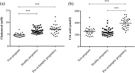
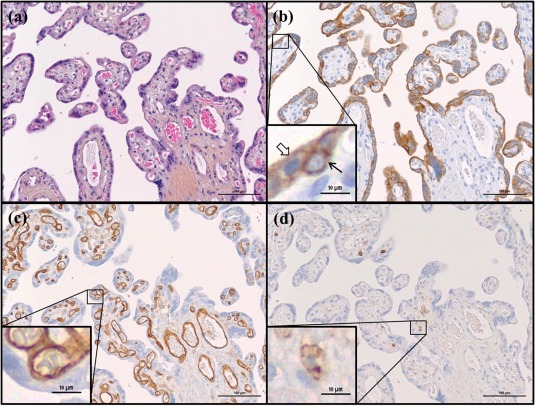
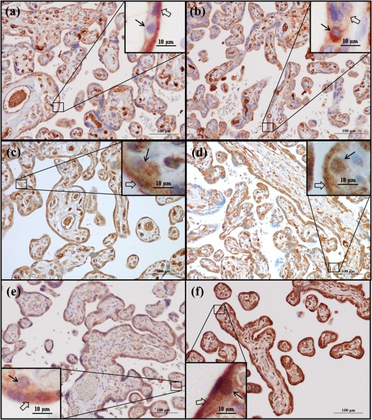
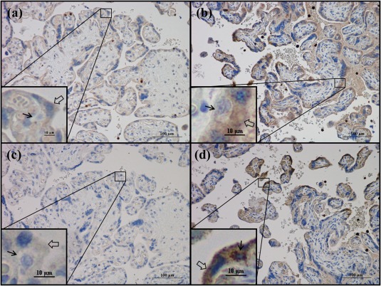
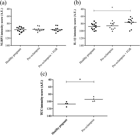
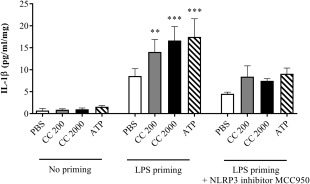
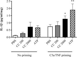
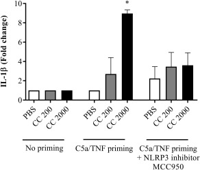
Pre-eclampsia is associated with increased levels of cholesterol and uric acid and an inflamed placenta expressing danger-sensing pattern recognition receptors (PRRs). Crystalline cholesterol and uric acid activate the PRR Nod-like receptor protein (NLRP)3 inflammasome to release interleukin (IL)-1β and result in vigorous inflammation. We aimed to characterize crystal-induced NLRP3 activation in placental inflammation and examine its role in pre-eclampsia. We confirmed that serum total cholesterol and uric acid were elevated in pre-eclamptic compared to healthy pregnancies and correlated positively to high sensitivity C-reactive protein (hsCRP) and the pre-eclampsia marker soluble fms-like tyrosine kinase-1 (sFlt-1). The NLRP3 inflammasome pathway components (NLRP3, caspase-1, IL-1β) and priming factors [complement component 5a (C5a) and terminal complement complex (TCC)] were co-expressed by the syncytiotrophoblast layer which covers the placental surface and interacts with maternal blood. The expression of IL-1β and TCC was increased significantly and C5a-positive regions in the syncytiotrophoblast layer appeared more frequent in pre-eclamptic compared to normal pregnancies. In-vitro activation of placental explants and trophoblasts confirmed NLRP3 inflammasome pathway functionality by complement-primed crystal-induced release of IL-1β. This study confirms crystal-induced NLRP3 inflammasome activation located at the syncytiotrophoblast layer as a mechanism of placental inflammation and suggests contribution of enhanced NLRP3 activation to the harmful placental inflammation in pre-eclampsia.

Keywords: NLRP3; cholesterol; inflammation; placenta; pre-eclampsia.


Figures

Figure 1
Figure 1
Maternal serum levels of total cholesterol and uric acid. Circulating levels of (a) total cholesterol and (b) uric acid were measured in serum from non‐pregnant (n = 28), healthy (n = 43) and pre‐eclamptic (n = 34) pregnant women. Data were analysed using the Kruskal–Wallis test with Dunn's multiple comparison post‐hoc test. ***P < 0·0001.
Figure 2
Figure 2
Haematoxylin erythrosine saffron (HES) staining and cell type markers in third trimester placenta. Representative images of healthy placental tissue at gestational age 38 weeks are shown stained by (a) HES and immunohistochemical staining of (b) the trophoblast marker cytokeratin 7 (CK7), (c) the endothelium marker CD31 and (d) the leucocyte marker CD45. Black arrow indicates cytotrophoblast, transparent arrow indicates syncytiotrophoblast.
Figure 3
Figure 3
Nod‐like receptor protein 3 (NLRP3), caspase‐1 and interleukin (IL)‐1β protein expression in third‐trimester placenta. Immunohistochemical staining of (a,b) Nod‐like receptor protein 3 (NLRP3), (c,d) caspase‐1 and (e,f) interleukin (IL)‐1β. Representative images of healthy placentas at gestational age (a,c) 40 + 0 and (e) 39 + 0 weeks, and pre‐eclamptic placentas at gestational age (b,d) 33 + 6 and (f) 33 + 5 weeks are shown. Black arrows indicate cytotrophoblasts, transparent arrows indicate syncytiotrophoblast.
Figure 4
Figure 4
Terminal complement complex (TCC) and complement component 5a (C5a) protein expression in third‐trimester placenta. Immunohistochemical staining of (a,b) TCC and (c,d) C5a. Representative images of (a,c) a healthy placenta at gestational age 40 + 0 and (b,d) a pre‐eclamptic placenta at gestational age 29 + 6 weeks are shown. Black arrows indicate cytotrophoblasts, transparent arrows indicate syncytiotrophoblast.
Figure 5
Figure 5
Protein expression levels of Nod‐like receptor protein 3 (NLRP3), interleukin (IL)‐1β and terminal complement complex (TCC) in third‐trimester placenta. (a,b) For NLRP3 and IL‐1β the syncytiotrophoblast cytoplasmic staining was quantified in healthy (n = 13) and pre‐eclamptic placentas without (n = 11) and with (n = 12) fetal growth restriction (FGR), and data were analysed using one‐way analysis of variance (anova) with Tukey's multiple comparison post‐hoc test. (c) TCC expression level was quantified in the syncytiotrophoblast cytoplasm of healthy (n = 4) and pre‐eclamptic placentas (n = 4), and data were analysed using the two‐tailed Mann–Whitney test. *P < 0·05. AU = arbitrary units.
Figure 6
Figure 6
Interleukin (IL)‐1β response following cholesterol crystal stimulation of lipopolysaccharide (LPS)‐primed placental explants, and the effect of Nod‐like receptor protein 3 (NLRP3) inhibition. Third‐trimester chorionic villous explants from six healthy pregnancies were first incubated with or without LPS, before adding cholesterol crystals (CC, 200 or 2000 µg/ml) or adenosine triphosphate (ATP). Three biological replicates were also treated with the NLRP3 inflammasome inhibitor MCC950. Six technical replicates were included for each experimental condition. Release of IL‐1β was measured by enzyme‐linked immunosorbent assay (ELISA), and is presented as mean ± standard error of the mean (s.e.m.) relative to explant weight. Data were analysed using the Kruskal–Wallis test with Dunn's multiple comparison post‐hoc test. *P < 0·05; **P < 0·01; ***P < 0·001. Lactate dehydrogenase viability analysis showed no toxic effect on the placental cells (Supporting information, Fig. S3a). PBS = phosphate‐buffered saline.
Figure 7
Figure 7
Interleukin (IL)‐1β response following C5a/tumour necrosis factor (TNF)‐α primed cholesterol crystal stimulation of placental explants. Third‐trimester chorionic villous explants from four healthy pregnancies were incubated with or without complement component 5a (C5a) and TNF‐α before stimulation with cholesterol crystals (CC, 200 or 2000 µg/ml) or adenosine triphosphate (ATP). Six technical replicates were included for each experimental condition. The level of IL‐1β was measured in supernatant using enzyme‐linked immunosorbent assay (ELISA) and presented as mean ± standard error of the mean (s.e.m.) relative to explant weight. Data were analysed using the Kruskal–Wallis test with Dunn's multiple comparison post‐hoc test. *P < 0·05; **P < 0·01. Lactate dehydrogenase viability analysis showed no toxic effect on the placental cells (Supporting information, Fig. S3b). PBS = phosphate‐buffered saline.
Figure 8
Figure 8
Interleukin (IL)‐1β response following complement component 5a (C5a)/tumour necrosis factor (TNF)‐primed cholesterol crystal stimulation of trophoblasts. The trophoblast cell line SGHPL‐5 was incubated with or without C5a and TNF‐α before stimulation with cholesterol crystals (CC, 200 or 2000 µg/ml). Treatment with the Nod‐like receptor protein 3 (NLRP3) inflammasome inhibitor MCC950 was included. IL‐1β release was measured by enzyme‐linked immunosorbent assay (ELISA) and presented as fold change of triplicates from three independent experiments. Data were analysed using the Kruskal–Wallis test with Dunn's multiple comparison post‐hoc test. *P < 0·05. Lactate dehydrogenase viability analysis showed no toxic effect on the cells (Supporting information, Fig. S4). PBS = phosphate‐buffered saline.

References

    1. Watts DH, Krohn MA, Wener MH, Eschenbach DA. C‐reactive protein in normal pregnancy. Obstet Gynecol 1991; 77:176–80. - PubMed
    1. Redman CW, Sargent IL. Preeclampsia and the systemic inflammatory response. Semin Nephrol 2004; 24:565–70. - PubMed
    1. Kalinderis M, Papanikolaou A, Kalinderi K et al Elevated serum levels of interleukin‐6, interleukin‐1beta and human chorionic gonadotropin in pre‐eclampsia. Am J Reprod Immunol 2011; 66:468–75. - PubMed
    1. Ernst GD, de Jonge LL, Hofman A et al C‐reactive protein levels in early pregnancy, fetal growth patterns, and the risk for neonatal complications: the generation R study. Am J Obstet Gynecol 2011; 205:132.e1–12. - PubMed
    1. Duley L. The global impact of pre‐eclampsia and eclampsia. Semin Perinatol 2009; 33:130–7. - PubMed

Publication types

MeSH terms