. 2018 Sep;57(9):1130-1143.
doi: 10.1002/mc.22830. Epub 2018 May 8.

A novel tricarbonylmethane agent (CMC2.24) reduces human pancreatic tumor growth in mice by targeting Ras

Affiliations

A novel tricarbonylmethane agent (CMC2.24) reduces human pancreatic tumor growth in mice by targeting Ras

Naveen A Mallangada et al. Mol Carcinog. 2018 Sep.

Abstract

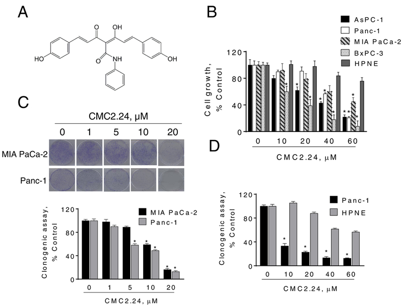
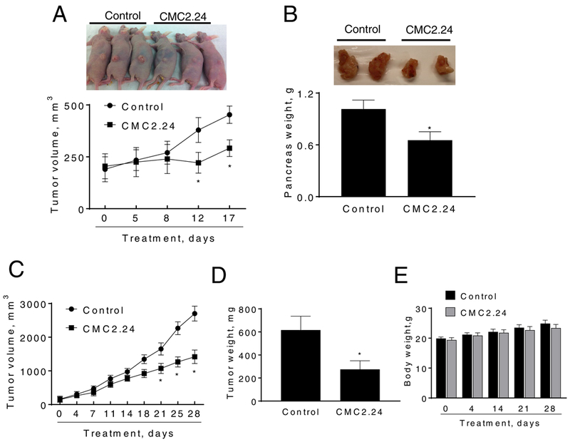
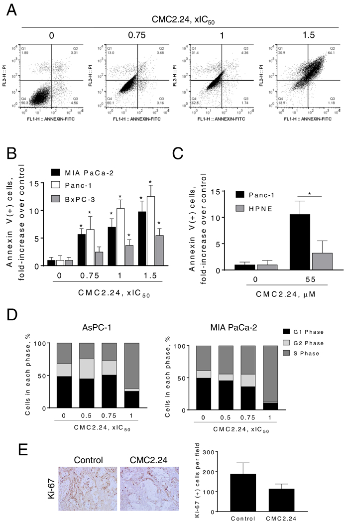
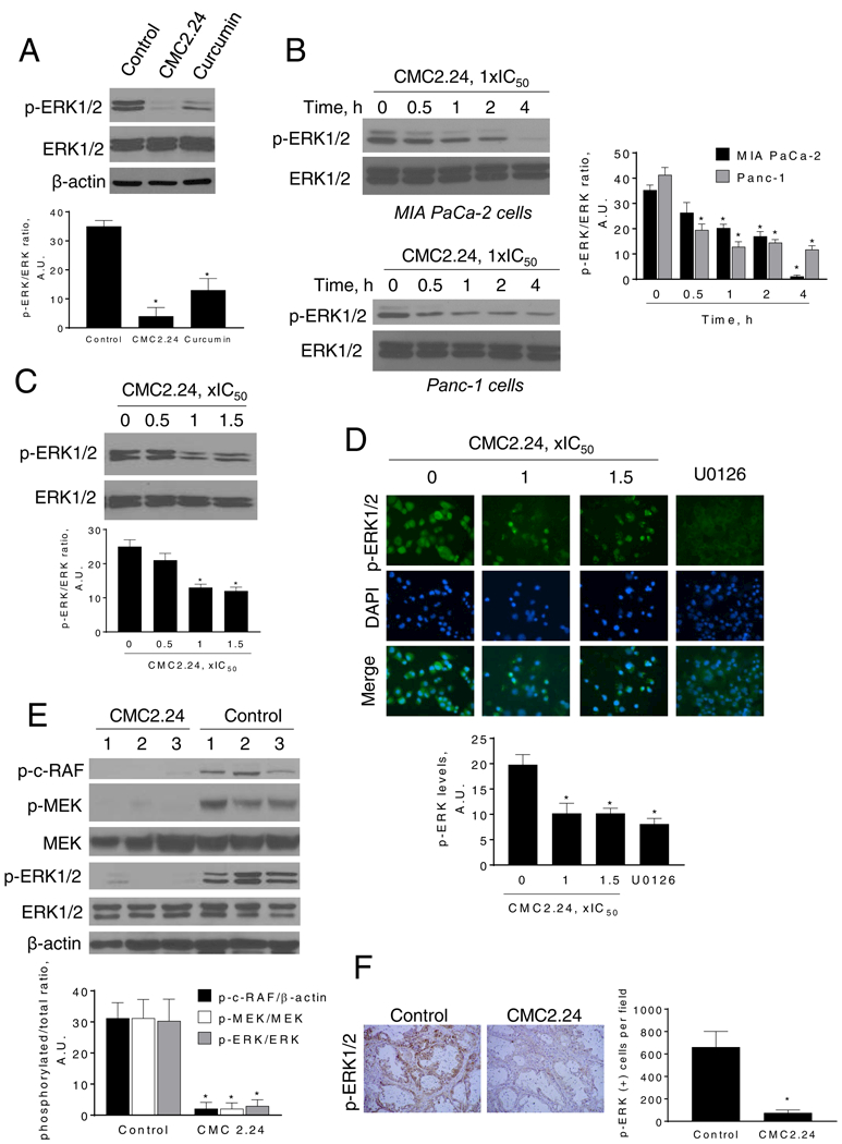
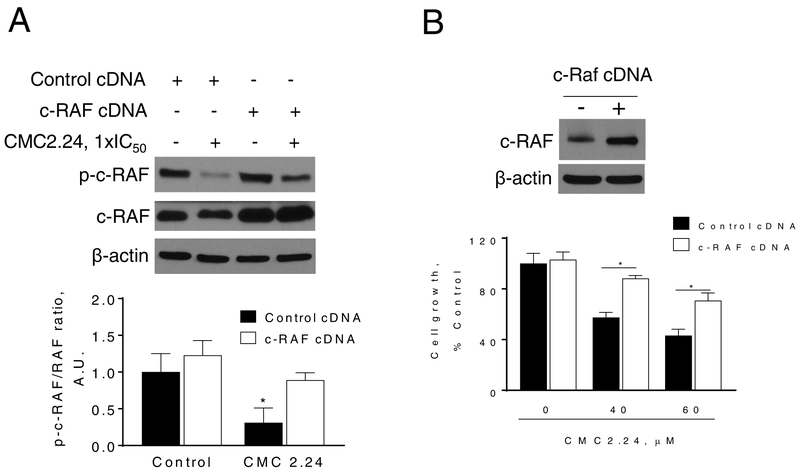
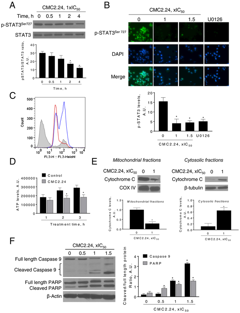
Pancreatic Cancer (PC) is a deadly disease in need of new therapeutic options. We recently developed a novel tricarbonylmethane agent (CMC2.24) as a therapeutic agent for PC, and evaluated its efficacy in preclinical models of PC. CMC2.24 inhibited the growth of various human PC cell lines in a concentration and time-dependent manner. Normal human pancreatic epithelial cells were resistant to CMC2.24, indicating selectivity. CMC2.24 reduced the growth of subcutaneous and orthotopic PC xenografts in mice by up to 65% (P < 0.02), and the growth of a human patient-derived tumor xenograft by 47.5% (P < 0.03 vs vehicle control). Mechanistically, CMC2.24 inhibited the Ras-RAF-MEK-ERK pathway. Based on Ras Pull-Down Assays, CMC2.24 inhibited Ras-GTP, the active form of Ras, in MIA PaCa-2 cells and in pancreatic acinar explants isolated from Kras mutant mice, by 90.3% and 89.1%, respectively (P < 0.01, for both). The inhibition of active Ras led to an inhibition of c-RAF, MEK, and ERK phosphorylation by 93%, 91%, and 87%, respectively (P < 0.02, for all) in PC xenografts. Furthermore, c-RAF overexpression partially rescued MIA PaCa-2 cells from the cell growth inhibition by CMC2.24. In addition, downstream of ERK, CMC2.24 inhibited STAT3 phosphorylation levels at the serine 727 residue, enhanced the levels of superoxide anion in mitochondria, and induced intrinsic apoptosis as shown by the release of cytochrome c from the mitochondria to the cytosol and the further cleavage of caspase 9 in PC cells. In conclusion, CMC2.24, a potential Ras inhibitor, is an efficacious agent for PC treatment in preclinical models, deserving further evaluation.

Keywords: CMC2.24; ERK; Kras; Ras; curcumin; pancreatic cancer.


Conflict of interest statement

Conflict of interest disclosure: LMG and FJ are listed as inventors on several issued patents on chemically-modified curcumins & these have been assigned to Stony Brook University (LMG) and to Chem-Master, Int (FJ). Moreover, both have an equity position in Traverse Biosciences, Inc, the company that provided the test compound. All other authors declare no conflict of interest.

Figures

Figure 1:
Figure 1:. CMC2.24 inhibits pancreatic cancer growth in vitro.
A: Chemical structure of the novel agent CMC2.24 (TRB-N0224). B: CMC2.24 inhibits PC cell growth in a concentration-dependent manner. Cell growth was determined in AsPC-1, Panc-1, MIA PaCa-2 and BxPC-3 PC cells, as well as in the human pancreatic normal epithelial cells (HPNE) after treatment with escalating concentrations of CMC2.24 for 24 h. Results are expressed as % control. (*p < 0.05, vs. control). C: CMC2.24 inhibits PC cell colony formation in a concentration-dependent manner in MIA PaCa-2 and Panc-1 cells. (*p < 0.05, vs. control). D: Colony formation assay in Panc-1 and HPNE cells. Results are expressed as the mean ± SEM (*p < 0.05, vs. control).
Figure 2:
Figure 2:. CMC2.24 inhibits the growth of pancreatic cancer xenografts.
A: CMC2.24 inhibits Panc-1 tumor growth in nude mice. Panc-1 cells were injected subcutaneously in the flank areas of nude mice and when palpable tumors were observed, the mice received CMC2.24 50 mg/kg/d by oral gavage in corn oil or just corn oil (control) for 17 days. B: Mouse KPC cells were orthotopically implanted in immunocompetent mice, which were then treated without (control) or with CMC2.24 50 mg/kg for 9 days. Pancreas weight at sacrifice. Results are expressed as the mean ± SEM (*p < 0.05, vs. control). C: CMC2.24 reduces the growth of a PDTX in SCID mice. D: Tumor weight at sacrifice. Results are expressed as the mean ± SEM (*p < 0.05, vs. control). E: Mice body weight over time for control and CMC2.24 treated groups. Results are expressed as the mean ± SEM.
Figure 3:
Figure 3:. The cell kinetic effect of CMC2.24 in pancreatic cancer cells.
A: AsPC-1 cells treated with CMC2.24 for 24 h were stained with Annexin V/propidium iodide, and the percentage of apoptotic cells was determined by flow cytometry. B: MIA Paca-2, Panc-1 and BxPC-3 cells treated with various concentrations of CMC2.24 for 24 h were stained with Annexin V/propidium iodide, and the percentage of apoptotic cells was determined by flow cytometry. Results are expressed as fold-increase compared with the percentages of apoptotic cells in the control cells. *p<0.05, vs. control. C: Differential cytotoxic effect of CMC2.24 in Panc-1 cells compared with that in HPNE. Apoptosis was determined by flow cytometry in HPNE and Panc-1 cells incubated with or without 55 μM of CMC2.24 for 24h. Results are expressed as fold-increase compared with the percentages of apoptotic cells in the control cells. D: CMC blocks the S/G2 cell cycle phase transition after 24 h treatment in human AsPC-1 and MIA PaCa-2 cells, determined by flow cytometry following propidium iodide staining. E: The percentage of proliferating cells in vehicle or CMC2.24-treated PDTX xenografts were determined by Ki-67 staining. Representative images (x20) of tissue sections from PDTX tumors treated with either vehicle (control) or CMC2.24 and stained for Ki-67 expression (proliferation marker). The proliferation indices of xenograft tumors were determined and expressed as the mean ± SEM.
Figure 4:
Figure 4:. CMC2.24 inhibits ERK1/2 activation in pancreatic cancer cells and xenografts.
A: Immunoblots of p-ERK and ERK in MIA PaCa-2 cells treated with CMC2.24 or curcumin at 1xIC50 for 4 h. Results we quantified as the ration between p-ERK/ERK; *p<0.05 versus control. B: Immunoblots of p-ERK and ERK in MIA PaCa-2 and Panc-1 total cell extracts treated with CMC2.24 1xIC50 for up to 4 h. Bands were quantified and results expressed as the p-ERK/ERK ratio. *p<0.05 versus time zero. C: Immunoblots of p-ERK and ERK in mouse KPC cells treated with CMC2.24 at 0.5x, 1x or 1.5x IC50 and quantified p-ERK values. *p<0.05 versus control. D: Immunofluorescence staining for p-ERK1/2 in Panc-1 cells treated with CMC2.24 (1x and 1.5xIC50) or U0126 (10 μM) for 3 h. Representative images in each group from three independent experiments are shown (original magnification, x20). The immunofluorescence intensity in each group was quantified. *p<0.05 versus control. E: Panc-1 xenograft tumor lysates were analyzed for p-c-RAF, p-MEK, MEK, pERK1/2 and ERK1/2 by immunoblotting. Loading control: β-actin. Each lane represents a different tumor sample. Bands were quantified and results are expressed as the ratio of phospho/total expression levels for each protein; *p<0.05 vs. control. F: Representative images (x20) of tissue sections from PDTX tumors treated with either vehicle (control) or CMC2.24 and stained for p-ERK1/2. The percentage of p-ERK1/2-positive cells per field was determined and expressed as mean ± SEM. *p <0.01 vs. control.
Figure 5:
Figure 5:. CMC inhibits Ras activation in pancreatic cancer.
A: Immunoblots of Ras-GTP (active-Ras) and total K-Ras in MIA PaCa-2 cells treated without or with CMC2.24, as indicated, for 3 h. B: Immunoblots of Ras-GTP (active Ras) in cell protein extracts from primary acinar explants isolated from Kras-active mice treated with CMC2.24 1x and 1.5xIC50, for 3 h. *p <0.05 vs. control.
Figure 6:
Figure 6:. c-RAF overexpression ameliorates, in part, the cell growth inhibition by CMC2.24.
MIA PaCa-2 cells were transfected with a control (cDNA) or c-RAF-expressing plasmid for 48 h and then treated with CMC2.24 for 24 h. A: Immunoblots of p-c-RAF and c-RAF in MIA PaCa-2 total cell extracts treated with CMC2.24 1xIC50 for 4 h. Bands were quantified and results expressed as the p-c-RAF/c-RAF ratio. *p<0.05 versus control. B: Cell growth was evaluated by the MTT assay. c-RAF overexpression rescues cells from the cell growth inhibition induced by CMC2.24. Top: c-RAF expression status in whole cell protein lysates following transfection.
Figure 7:
Figure 7:. CMC2.24 reduces STAT3Ser727 phosphorylation levels, induces mitochondrial reactive oxygen species and mitochondrial cell death in pancreatic cancer cells.
A: Immunoblots of STAT3 and phosphorylated STAT3 (STAT3Ser727) from Panc-1 cells treated with CMC2.24 for up to 4 h. Bands were quantified and results expressed as the p-STAT3Ser727/STAT3 ratio. *p<0.05 versus time zero. B: Immunofluorescence staining for p-STAT3Ser727 in Panc-1 cells treated with CMC2.24 (1x and 1.5xIC50) or U0126 (10 μM) for 3 h. Representative images in each group from three independent experiments are shown (original magnification, x20). The immunofluorescence intensity in each group was quantified. *p<0.05 versus control. C: MitoSOX Red fluorescence was measured by flow cytometry in MIA PaCa-2 cells treated with CMC2.24 for 3 h. MitoSOX Red fluorescent histograms for control (grey), CMC2.24 1xIC50 (red line) and CMC2.24 1.5xIC50 (blue line) are shown. D: ATP levels were determined in MIA PaCa-2 cells treated without (control) or with (CMC2.24 1xIC50) for up to 3 h. Results were quantified and expressed as mean ± SEM; *p<0.05 vs. control. E: CMC induces the release to the cytosol of cytochrome C. Mitochondrial fractions loading control: COX IV. Cytosolic fractions loading control: β-tubulin. Bands were quantified and results are shown as mean ± SEM; *p<0.05 vs. control. F: Immunoblots for full length and cleaved caspase 9 as well as full length and cleaved PARP in total cell protein extracts from MIA PaCa-2 cells treated with CMC2.24, as indicated, for 24 h. Loading control: β-actin. Bands were quantified and results are shown as the ratio between the cleaved/full length protein; *p<0.05 vs. control.

Similar articles

Cited by

References

    1. Rahib L, Smith BD, Aizenberg R, Rosenzweig AB, Fleshman JM, Matrisian LM. Projecting cancer incidence and deaths to 2030: the unexpected burden of thyroid, liver, and pancreas cancers in the United States. Cancer Res 2014;74(11):2913–2921. - PubMed
    1. Kleeff J, Korc M, Apte M et al. Pancreatic cancer. Nature reviews Disease primers 2016;2:16022. - PubMed
    1. Ying JE, Zhu LM, Liu BX. Developments in metastatic pancreatic cancer: is gemcitabine still the standard? World journal of gastroenterology : WJG 2012;18(8):736–745. - PMC - PubMed
    1. Hruban RH, van Mansfeld AD, Offerhaus GJ et al. K-ras oncogene activation in adenocarcinoma of the human pancreas. A study of 82 carcinomas using a combination of mutant-enriched polymerase chain reaction analysis and allele-specific oligonucleotide hybridization. The American journal of pathology 1993;143(2):545–554. - PMC - PubMed
    1. di Magliano MP, Logsdon CD. Roles for KRAS in Pancreatic Tumor Development and Progression. Gastroenterology 2013;144(6):1220–1229. - PMC - PubMed

Publication types

MeSH terms

Substances