

Isosteviol sodium injection improves outcomes by modulating TLRs/NF-κB-dependent inflammatory responses following experimental traumatic brain injury in rats
- PMID: 29683870
- PMCID: PMC5999382
- DOI: 10.1097/WNR.0000000000001033
Isosteviol sodium injection improves outcomes by modulating TLRs/NF-κB-dependent inflammatory responses following experimental traumatic brain injury in rats
Abstract
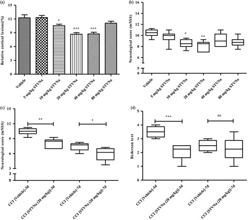
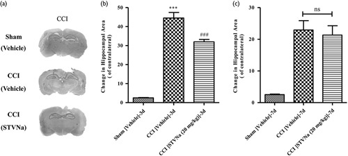
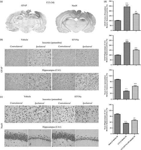
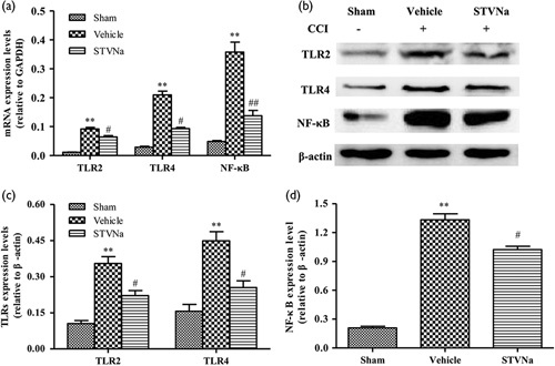
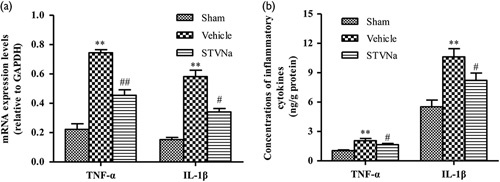
Previous studies have shown that isosteviol sodium (STVNa) protects against permanent cerebral ischemia injury by inhibition of the nuclear factor kappa-light-chain-enhancer of activated B cells (NF-κB)-mediated inflammatory responses. Overwhelming evidence shows that toll-like receptors (TLRs) are the upstream regulators of NF-κB. On the basis of the similarity of the pathology caused by traumatic brain injury (TBI) and stroke, we speculated that STVNa may have a therapeutic effect against TBI through regulation of the TLRs/NF-κB signaling-mediated inflammatory response. Thus, we studied the potential therapeutic effects of STVNa and the underlying mechanisms. Male rats, subjected to controlled cortical impact (CCI) injury, were injected intraperitoneally with STVNa (5, 10, 20, 40, and 80 mg/kg, daily for 3 or 7 days) after trauma. Neurobehavioral scores, relative numbers of cortical lesions, and histology were examined. We also measured the mRNA and protein expression levels of TLRs/NF-κB signaling pathway-related genes including TLR2, TLR4, and NF-κB by quantitative real-time-PCR and western blotting, respectively, and concentrations of tumor necrosis factor-α and interleukin-1β by an enzyme-linked immunosorbent assay. The results indicated that STVNa (20 mg/kg) showed significant neuroprotective effects 3 and 7 days after TBI, including the reduction of cortical lesions, improvement of the neurological severity score, significantly increased number of restored neurons, decreased number of astrocytes, and lower concentrations of tumor necrosis factor-α and interleukin-1β. Results from quantitative real-time-PCR and western blotting also show that the mRNA and protein expression levels of TLR2, TLR4, and NF-κB were significantly lower in STVNa-treated rats compared with the vehicle-treated rats. The administration of STVNa attenuates the TLR/NF-κB signaling pathway-mediated inflammatory responses in the injured rat brain, and this may be the mechanism by which STVNa improves the outcome following TBI.
Figures
Similar articles
-
Isosteviol Sodium Protects Against Permanent Cerebral Ischemia Injury in Mice via Inhibition of NF-κB-Mediated Inflammatory and Apoptotic Responses.J Stroke Cerebrovasc Dis. 2017 Nov;26(11):2603-2614. doi: 10.1016/j.jstrokecerebrovasdis.2017.06.023. Epub 2017 Aug 4. J Stroke Cerebrovasc Dis. 2017. PMID: 28784277
-
Progesterone administration modulates TLRs/NF-kappaB signaling pathway in rat brain after cortical contusion.Ann Clin Lab Sci. 2008 Winter;38(1):65-74. Ann Clin Lab Sci. 2008. PMID: 18316784
-
Omega-3 polyunsaturated fatty acid supplementation attenuates microglial-induced inflammation by inhibiting the HMGB1/TLR4/NF-κB pathway following experimental traumatic brain injury.J Neuroinflammation. 2017 Jul 24;14(1):143. doi: 10.1186/s12974-017-0917-3. J Neuroinflammation. 2017. PMID: 28738820 Free PMC article.
-
Pharmacological Effects of Polyphenol Phytochemicals on the Intestinal Inflammation via Targeting TLR4/NF-κB Signaling Pathway.Int J Mol Sci. 2022 Jun 22;23(13):6939. doi: 10.3390/ijms23136939. Int J Mol Sci. 2022. PMID: 35805952 Free PMC article. Review.
-
Therapeutic Correlation of TLR-4 Mediated NF-κB Inflammatory Pathways in Ischemic Injuries.Curr Drug Targets. 2024;25(15):1027-1040. doi: 10.2174/0113894501322228240830063605. Curr Drug Targets. 2024. PMID: 39279711 Review.
Cited by
-
Role of astroglial toll-like receptors (TLRs) in central nervous system infections, injury and neurodegenerative diseases.Brain Behav Immun. 2021 Jan;91:740-755. doi: 10.1016/j.bbi.2020.10.007. Epub 2020 Oct 8. Brain Behav Immun. 2021. PMID: 33039660 Free PMC article. Review.
-
Isosteviol Sodium Promotes Neurological Function Recovery in a Model of Spinal Cord Injury in Rats.Immun Inflamm Dis. 2025 Jan;13(1):e70110. doi: 10.1002/iid3.70110. Immun Inflamm Dis. 2025. PMID: 39783228 Free PMC article.
-
Neuroprotective Effects of Isosteviol Sodium in Murine Brain Capillary Cerebellar Endothelial Cells (cerebEND) After Hypoxia.Front Cell Neurosci. 2020 Oct 28;14:573950. doi: 10.3389/fncel.2020.573950. eCollection 2020. Front Cell Neurosci. 2020. PMID: 33192319 Free PMC article.
-
Isosteviol prevents the development of isoprenaline‑induced myocardial hypertrophy.Int J Mol Med. 2019 Nov;44(5):1932-1942. doi: 10.3892/ijmm.2019.4342. Epub 2019 Sep 17. Int J Mol Med. 2019. PMID: 31545484 Free PMC article.
-
Toll-Like Receptor Signaling Pathways: Novel Therapeutic Targets for Cerebrovascular Disorders.Int J Mol Sci. 2021 Jun 7;22(11):6153. doi: 10.3390/ijms22116153. Int J Mol Sci. 2021. PMID: 34200356 Free PMC article. Review.
References
-
- Runyan DK. The challenges of assessing the incidence of inflicted traumatic brain injury: a world perspective. Am J Prev Med 2008; 34:S112–S115. - PubMed
-
- Coronado VG, Xu L, Basavaraju SV, McGuire LC, Wald MM, Faul MD, et al. Surveillance for traumatic brain injury-related deaths – United States, 1997–2007. Morbidity and mortality weekly report. MMWR Surveill Summ 2011; 60:1–32. - PubMed
-
- Mcgarry LJ, Thompson D, Millham FH, Cowell L, Snyder PJ, Lenderking WR, Weinstein MC. Outcomes and costs of acute treatment of traumatic brain injury. J Trauma 2002; 53:1152. - PubMed
-
- Helmy A, De Simoni MG, Guilfoyle MR, Carpenter KL, Hutchinson PJ. Cytokines and innate inflammation in the pathogenesis of human traumatic brain injury. Prog Neurobiol 2011; 95:352–372. - PubMed
Publication types
MeSH terms
Substances
LinkOut - more resources
Full Text Sources
Other Literature Sources
Medical
