. 2018 Apr 23;13(4):e0196080.
doi: 10.1371/journal.pone.0196080. eCollection 2018.

Murine embryos exposed to human endometrial MSCs-derived extracellular vesicles exhibit higher VEGF/PDGF AA release, increased blastomere count and hatching rates

Affiliations

Murine embryos exposed to human endometrial MSCs-derived extracellular vesicles exhibit higher VEGF/PDGF AA release, increased blastomere count and hatching rates

Rebeca Blázquez et al. PLoS One. .

Abstract

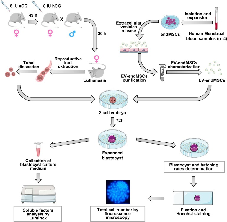
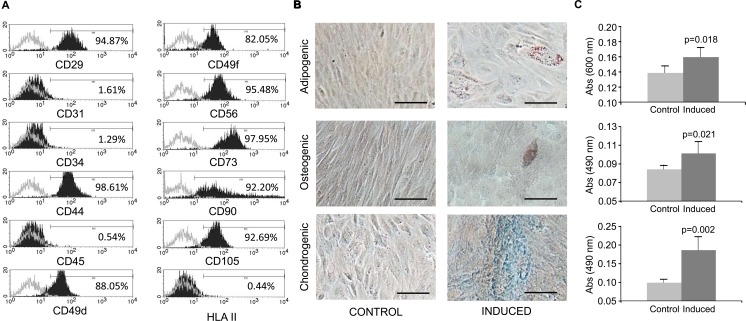
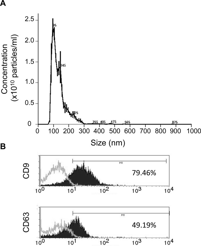
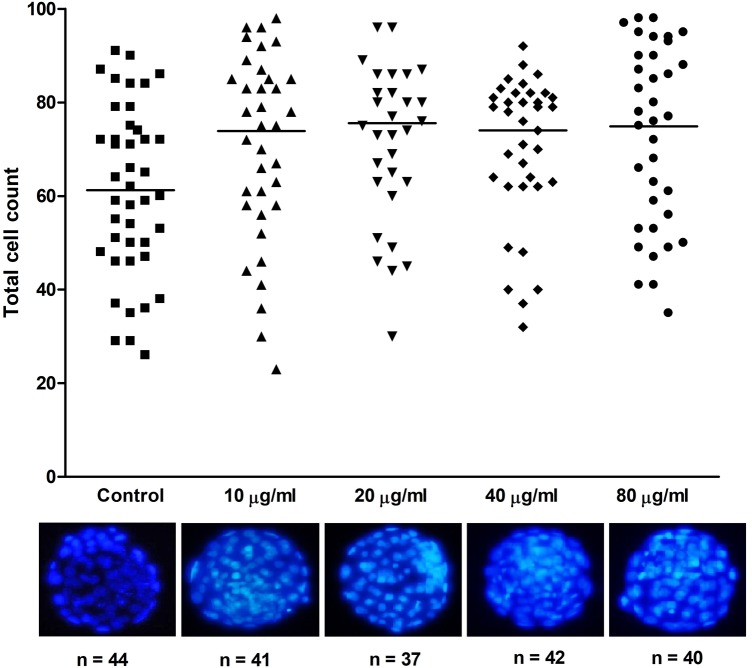
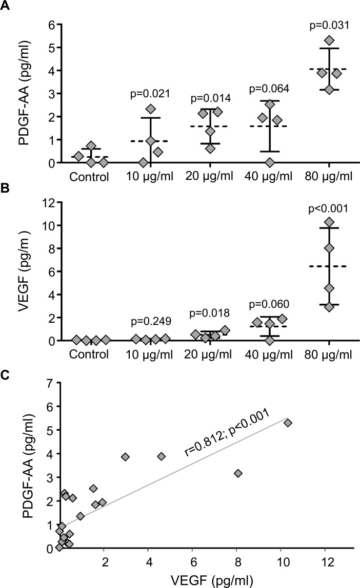
Endometrial Mesenchymal Stromal Cells (endMSCs) are multipotent cells with immunomodulatory and pro-regenerative activity which is mainly mediated by a paracrine effect. The exosomes released by MSCs have become a promising therapeutic tool for the treatment of immune-mediated diseases. More specifically, extracellular vesicles derived from endMSCs (EV-endMSCs) have demonstrated a cardioprotective effect through the release of anti-apoptotic and pro-angiogenic factors. Here we hypothesize that EV-endMSCs may be used as a co-adjuvant to improve in vitro fertilization outcomes and embryo quality. Firstly, endMSCs and EV-endMSCs were isolated and phenotypically characterized for in vitro assays. Then, in vitro studies were performed on murine embryos co-cultured with EV-endMSCs at different concentrations. Our results firstly demonstrated a significant increase on the total blastomere count of expanded murine blastocysts. Moreover, EV-endMSCs triggered the release of pro-angiogenic molecules from embryos demonstrating an EV-endMSCs concentration-dependent increase of VEGF and PDGF-AA. The release of VEGF and PDGF-AA by the embryos may indicate that the beneficial effect of EV-endMSCs could be mediating not only an increase in the blastocyst's total cell number, but also may promote endometrial angiogenesis, vascularization, differentiation and tissue remodeling. In summary, these results could be relevant for assisted reproduction being the first report describing the beneficial effect of human EV-endMSCs on embryo development.


Conflict of interest statement

Competing Interests: The authors have declared that no competing interests exist.

Figures

Fig 1
Fig 1. Description of the study and methodological design.
Fig 2
Fig 2. Phenotypical characterization and differentiation potential of endMSCs.
(A) Flow cytometry analysis of endMSCs isolated from menstrual blood. A representative histogram together with the expression levels is shown. The expression of cell surface markers is represented as the percentage of positive cells (black lined histogram) above the negative control (grey lined histogram). (B) In vitro differentiation potential of endMSCs. The cells were in vitro cultured for 21 days with standard medium (Control) or with specific differentiation media for adipogenic, osteogenic and chondrogenic lineages (Induced). Differentiation towards adipocytes, osteocytes and chondrocytes was evidenced by Oil Red O, Alizarin Red and Alcian Blue 8GX respectively. Scale bar: 100 μm. (C) The adipogenic (above), osteogenic (middle) and chondrogenic (below) differentiation degree was quantified by determining the absorbance of the extracts at 490 nm (Oil Red O and Alizarin Red S staining) and at 600 nm (Alcian Blue 8GX). Four independent experiments using four different cell lines were performed and a Mann-Whitney U test was used. p-values are shown in the figure.
Fig 3
Fig 3. Characterization of EV-endMSCs.
(A) Representative graph of nanoparticle tracking analysis to quantify size distribution and particle concentration of EV-endMSCs. (B) Flow cytometry analysis on EV-endMSCs for exosomal markers CD9 and CD63. A representative histogram together with the expression levels is shown. The expression of cell surface markers is represented as as the percentage of positive cells (black lined histogram) above the negative control (grey lined histogram).
Fig 4
Fig 4. Total cell number of murine blastocysts cultured in presence or absence of EV-endMSCs.
Total cell number of the obtained expanded blastocysts was obtained after Hoechst 33342 staining and subsequent evaluation by fluorescence microscopy. For each treatment, the individual blastomere count is represented. Horizontal bars show the mean values. All the treatments differ statistically from the control (p<0.05). Representative micrographs of expanded blastocysts cultured with varying dosages of EV-endMSCs are shown.
Fig 5
Fig 5. Quantification of VEGF and PDGF-AA secreted during embryo culture.
Soluble factors released by the developing embryos co-cultured with EV-endMSCS were quantified by the Luminex xMAP technology at the third day of embryo culture. (A) PDGF-AA secreted by blastocyst embryos. All the data were compared by Student t-test for paired comparisons with respect to control group. The mean (dotted line) ±SD from four independent experiments, as well as individual measures (rhombuses), are shown. (B) VEGF secreted by blastocyst embryos. All the data were compared by Student t-test for paired comparisons with respect to control group. The mean (dotted line) ±SD from four independent experiments, as well as individual measures (rhombuses), are shown. (C) Correlation between PDGF-AA and VEGF. Correlation line as well as individual measures (rhombuses) are represented. The Pearson correlation coefficient (r) together with its significance level (p) is shown.

References

    1. Khoury M, Alcayaga-Miranda F, Illanes SE, Figueroa FE. The promising potential of menstrual stem cells for antenatal diagnosis and cell therapy. Front Immunol. 2014;5: 205 doi: 10.3389/fimmu.2014.00205 - DOI - PMC - PubMed
    1. Uder C, Brückner S, Winkler S, Tautenhahn H-M, Christ B. Mammalian MSC from selected species: Features and applications. Cytometry A. 2018;93: 32–49. doi: 10.1002/cyto.a.23239 - DOI - PubMed
    1. Burke J, Hunter M, Kolhe R, Isales C, Hamrick M, Fulzele S. Therapeutic potential of mesenchymal stem cell based therapy for osteoarthritis. Clin Transl Med. 2016;5: 27 doi: 10.1186/s40169-016-0112-7 - DOI - PMC - PubMed
    1. Volarevic V, Nurkovic J, Arsenijevic N, Stojkovic M. Concise review: Therapeutic potential of mesenchymal stem cells for the treatment of acute liver failure and cirrhosis. Stem Cells. 2014;32: 2818–2823. doi: 10.1002/stem.1818 - DOI - PubMed
    1. Rani S, Ryan AE, Griffin MD, Ritter T. Mesenchymal Stem Cell-derived Extracellular Vesicles: Toward Cell-free Therapeutic Applications. Mol Ther. 2015;23: 812–823. doi: 10.1038/mt.2015.44 - DOI - PMC - PubMed

Publication types

MeSH terms

Substances