. 2018 Apr 23;14(4):e1006998.
doi: 10.1371/journal.ppat.1006998. eCollection 2018 Apr.

PgaB orthologues contain a glycoside hydrolase domain that cleaves deacetylated poly-β(1,6)-N-acetylglucosamine and can disrupt bacterial biofilms

Affiliations

PgaB orthologues contain a glycoside hydrolase domain that cleaves deacetylated poly-β(1,6)-N-acetylglucosamine and can disrupt bacterial biofilms

Dustin J Little et al. PLoS Pathog. .

Abstract

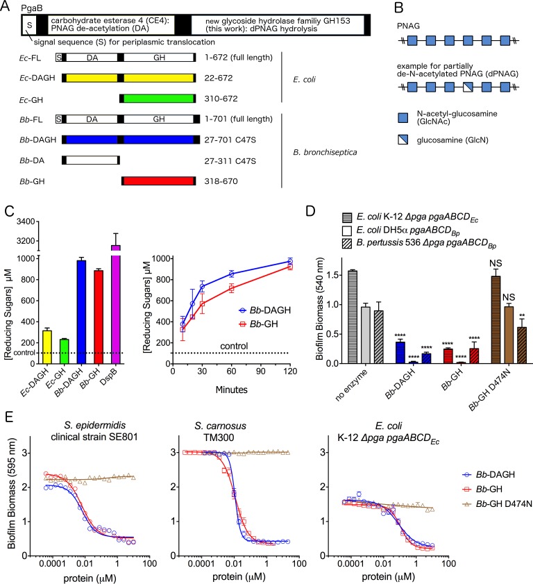
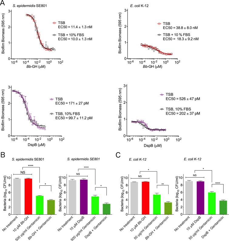
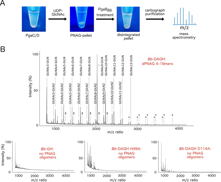
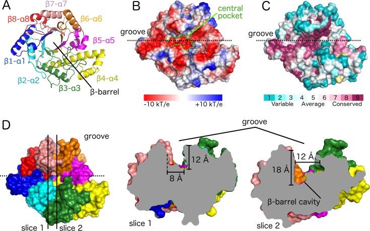
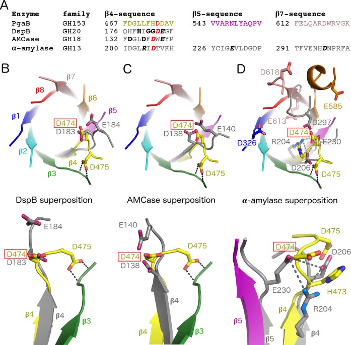
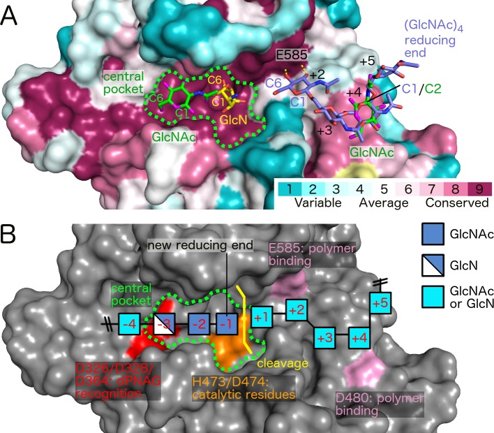
Poly-β(1,6)-N-acetyl-D-glucosamine (PNAG) is a major biofilm component of many pathogenic bacteria. The production, modification, and export of PNAG in Escherichia coli and Bordetella species require the protein products encoded by the pgaABCD operon. PgaB is a two-domain periplasmic protein that contains an N-terminal deacetylase domain and a C-terminal PNAG binding domain that is critical for export. However, the exact function of the PgaB C-terminal domain remains unclear. Herein, we show that the C-terminal domains of Bordetella bronchiseptica PgaB (PgaBBb) and E. coli PgaB (PgaBEc) function as glycoside hydrolases. These enzymes hydrolyze purified deacetylated PNAG (dPNAG) from Staphylococcus aureus, disrupt PNAG-dependent biofilms formed by Bordetella pertussis, Staphylococcus carnosus, Staphylococcus epidermidis, and E. coli, and potentiate bacterial killing by gentamicin. Furthermore, we found that PgaBBb was only able to hydrolyze PNAG produced in situ by the E. coli PgaCD synthase complex when an active deacetylase domain was present. Mass spectrometry analysis of the PgaB-hydrolyzed dPNAG substrate showed a GlcN-GlcNAc-GlcNAc motif at the new reducing end of detected fragments. Our 1.76 Å structure of the C-terminal domain of PgaBBb reveals a central cavity within an elongated surface groove that appears ideally suited to recognize the GlcN-GlcNAc-GlcNAc motif. The structure, in conjunction with molecular modeling and site directed mutagenesis led to the identification of the dPNAG binding subsites and D474 as the probable catalytic acid. This work expands the role of PgaB within the PNAG biosynthesis machinery, defines a new glycoside hydrolase family GH153, and identifies PgaB as a possible therapeutic agent for treating PNAG-dependent biofilm infections.


Conflict of interest statement

I have read the journal's policy and the authors of this manuscript have the following competing interests: A patent covering the use of the PgaB and other glycoside hydrolases for the prevention and/or disruption of biofilms has been filed (Application number WO2015/184526 A1).

Figures

Fig 1
Fig 1. PgaB exhibits glycoside hydrolase activity.
Schematic representation of the (A) PgaB constructs used in this study and (B) PNAG and dPNAG composition. (C) Reducing sugar assay with 2 mg/ml dPNAG purified from S. aureus with 2 μM PgaB variants and DspB over 24 h and PgaBBb hydrolase activity against dPNAG over 2 h. Error bars represent the standard error (S.E.) from two independent experiments performed in duplicate. (D) Biofilm disruption assay performed with 1.3 μM enzyme and the indicated strains. ****P ≤ 0.0001, **P ≤ 0.01, NS: no significant difference. Statistical significance was evaluated using two-way analysis of variance and Tukey’s multiple comparison test. (E) Dose response curves examining S. carnosus, S. epidermidis, and E. coli biofilm disruption. In panels D/E the error bars represent the S.E. and n = 3. In all graphs, Bb-DAGH is coloured blue, Bb-GH red, and the D474N variant brown.
Fig 2
Fig 2. Bb-GH is active in fetal bovine serum and potentiates killing by gentamicin.
(A) Dose response curves investigating the effect of fetal bovine serum on Bb-GH and DspB activity during disruption of S. epidermidis SE801 and E. coli K-12 biofilms. Error bars represent S.E., n = 3. (B) Enumeration of S. epidermidis SE801 after biofilm treatment with Bb-GH or DspB and 500 μg/ml gentamicin for 20 h. (C) Enumeration of E. coli K-12 after biofilm treatment with Bb-GH or DspB and 50 μg/ml gentamicin for 4 h. For (B) and (C) the mean was calculated from three independent experiments, error bars represent S.E.M. Statistical significance was calculated using one-way analysis of variance and Tukey’s multiple comparison test. ****P ≤ 0.0001, **P ≤ 0.01, *P ≤ 0.05, NS: no significant difference.
Fig 3
Fig 3. Bb-DAGH degrades in situ produced PNAG.
(A) Schematic of the in situ PNAG digestion assay. PNAG is produced in situ using PgaCD containing E. coli membranes. PNAG production and enzymatic cleavage is verified by mass-spectrometry after purification of the hydrolyzed sample. (B) MALDI-TOF MS profiles of product released by PgaBBb variants. The profile of the Bb-DAGH treated sample is enlarged to show details in the detected dPNAG/PNAG cleavage products. Profiles of samples containing no detectable dPNAG/PNAG components are displayed in the bottom row. Symbols represent identified structures less than 1% of relative intensity, ‡: fully N-acetylated structures, †: mono-deacetylated structures, *: di-deacetylated structures.
Fig 4
Fig 4. Structural analysis of Bb-DAGH treated PNAG reveals a consensus recognition motif.
(A) MALDI-TOF MS/MS profile of the m/z ion = 2031.71, a mono-deacetylated PNAG 10-mer. Oligosaccharides were reduced by NaBH4 allowing the reducing terminus to be defined. The asterisk indicates the reducing end of the molecule. (B) Graphical representation of the dPNAG 10-mer using the same key as depicted in Fig 1B. Sugar units are numbered relative to the cleavage site at the reducing end.
Fig 5
Fig 5. The (β/α)8 TIM-barrel of Bb-GH has a long and deep groove with a highly conserved central pocket.
(A) Cartoon representation of the (β/α)8 barrel with each β/α segment shown in a different color. (B) Electrostatic surface representation in the same orientation as panel A. The central pocket forming the deepest region of the groove is highlighted in green. (C) Surface representation with residues coloured based on conservation level (yellow: insufficient data). (D) Surface representation in the same colour coding and orientation as panel A. Slice 1 and 2 display different sections of the groove as indicated by the vertical lines in left panel and viewed from along the groove from left to right.
Fig 6
Fig 6. Residue D474 aligns with the catalytic aspartate from structurally similar GH families.
(A) Sequence comparison of regions containing catalytic residues. Residues in bold form the consensus motif for each GH family. Residues in bold italics are the catalytic residues. D474 and the structurally aligned aspartates are highlighted in red. Superposition of Bb-GH with (B) DspB (PDB 1YHT) [42], (C) AMCase (PDB 2YBU) [81], and (D) α-amylase (PDB 7TAA) [82]. Bb-GH β-strands are shown in the same color scheme as Fig 5A, all other enzymes are shown in grey.
Fig 7
Fig 7. Mutagenesis suggests D474 is involved in catalysis and other charged residues crucial for dPNAG binding.
(A) Reducing sugar assay with 2 mg/ml dPNAG purified from S. aureus with 2 μM PgaB constructs over 24 h. Error bars, S.E. from two independent experiments performed in duplicate. (B) E. coli biofilm disruption assay. Error bars represent the S.E. with n = 3. (C) EC50 values from E. coli biofilm disruption assay. ND: EC50 not determined as > 50% biomass remained or no plateau was reached after treatment with 5 μM enzyme for 2 h. Error bars show 95% confidence interval, and the same color scheme in A is used. (D) Transparent surface representation of the Bb-GH binding groove with mutated amino acids shown in stick representation. Note that D364 is fully buried and only visible due to the transparent representation.
Fig 8
Fig 8. Sequence and structural comparison of Ec-GH and Bb-GH reveal differences in active site accessibility.
(A) Sequence alignment between Bb-GH and Ec-GH showing identical and similar residues shaded and boxed in black, respectively. Secondary structure elements of Bb-GH are shown above the sequence alignment with the canonical (β/α)8 elements labeled. Residues forming the active site pocket and analyzed by mutagenesis are highlighted with the same color scheme as in Fig 7, with loops 3 and 7 highlighted by green boxes. The sequence alignment figure was generated using ESPript 3.0 [83]. (B) Structural superposition of Bb-GH and Ec-GH shown in cartoon representation. (C) Surface representation of Ec-GH and Bb-GH. In (B) and (C) loops 3 and 7 are highlighted in red and green, respectively.
Fig 9
Fig 9. Proposed recognition of dPNAG required for polymer cleavage by Bb-GH.
(A) Surface representation of Bb-GH with residues coloured based on conservation level (yellow: insufficient data). Shown in stick representation are GlcN (PDB 4P7N), GlcNAc (PDB ID 4P7Q), and (GlcNAc)4 (PDB ID 4P7R) that have been co-crystallized with Ec-GH and were modeled into the Bb-GH structure. (B) Proposed cleavage mechanism based on mutagenesis and mass spectrometry analysis. Crucial surfaces are highlighted in red, orange, and pink. Positions -4 to +5 denote expected binding sites of GlcN/GlcNAc units relative to the cleavage site. Positions +2 to +5 were taken directly from (GlcNAc) 4, position -1 coincides approximately with a GlcN monomer bound to PgaBEc (PDB 4P7N) and position -3 with a GlcNAc monomer bound to PgaBEc (PDB 4P7Q) [24].

References

    1. Mishra M, Parise G, Jackson KD, Wozniak DJ, Deora R. The BvgAS signal transduction system regulates biofilm development in Bordetella. J Bacteriol. 2005;187(4):1474–84. doi: 10.1128/JB.187.4.1474-1484.2005 ; PubMed Central PMCID: PMC545624. - DOI - PMC - PubMed
    1. Irie Y, Mattoo S, Yuk MH. The Bvg virulence control system regulates biofilm formation in Bordetella bronchiseptica. J Bacteriol. 2004;186(17):5692–8. doi: 10.1128/JB.186.17.5692-5698.2004 ; PubMed Central PMCID: PMC516841. - DOI - PMC - PubMed
    1. Sukumar N, Nicholson TL, Conover MS, Ganguly T, Deora R. Comparative analyses of a cystic fibrosis isolate of Bordetella bronchiseptica reveal differences in important pathogenic phenotypes. Infect Immun. 2014;82(4):1627–37. doi: 10.1128/IAI.01453-13 ; PubMed Central PMCID: PMC3993408. - DOI - PMC - PubMed
    1. Serra DO, Conover MS, Arnal L, Sloan GP, Rodriguez ME, Yantorno OM, et al. FHA-mediated cell-substrate and cell-cell adhesions are critical for Bordetella pertussis biofilm formation on abiotic surfaces and in the mouse nose and the trachea. PLoS One. 2011;6(12):e28811 Epub 2012/01/05. doi: 10.1371/journal.pone.0028811 PONE-D-11-15276 [pii]. ; PubMed Central PMCID: PMC3245231. - DOI - PMC - PubMed
    1. Conover MS, Mishra M, Deora R. Extracellular DNA is essential for maintaining Bordetella biofilm integrity on abiotic surfaces and in the upper respiratory tract of mice. PLoS One. 2011;6(2):e16861 Epub 2011/02/25. doi: 10.1371/journal.pone.0016861 . - DOI - PMC - PubMed

Publication types

MeSH terms

Substances

LinkOut - more resources