Review
. 2018 Jul:90:350-370.
doi: 10.1016/j.neubiorev.2018.04.015. Epub 2018 Apr 20.

White matter pathways and social cognition

Affiliations
Review

White matter pathways and social cognition

Yin Wang et al. Neurosci Biobehav Rev. 2018 Jul.

Abstract

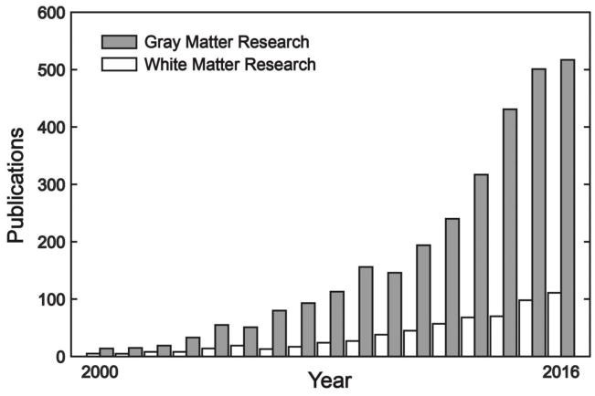
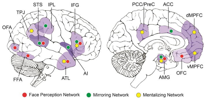
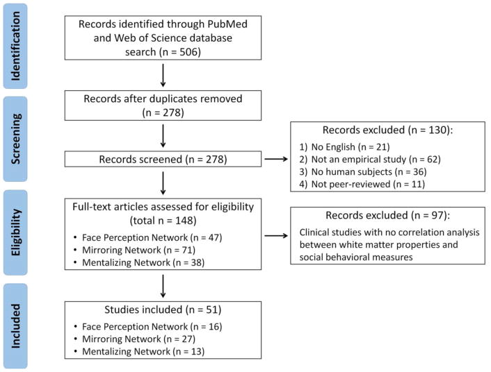
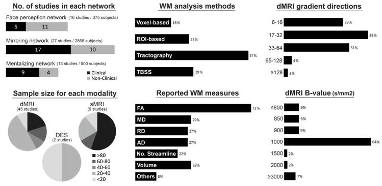
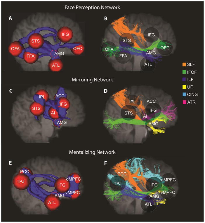
There is a growing consensus that social cognition and behavior emerge from interactions across distributed regions of the "social brain". Researchers have traditionally focused their attention on functional response properties of these gray matter networks and neglected the vital role of white matter connections in establishing such networks and their functions. In this article, we conduct a comprehensive review of prior research on structural connectivity in social neuroscience and highlight the importance of this literature in clarifying brain mechanisms of social cognition. We pay particular attention to three key social processes: face processing, embodied cognition, and theory of mind, and their respective underlying neural networks. To fully identify and characterize the anatomical architecture of these networks, we further implement probabilistic tractography on a large sample of diffusion-weighted imaging data. The combination of an in-depth literature review and the empirical investigation gives us an unprecedented, well-defined landscape of white matter pathways underlying major social brain networks. Finally, we discuss current problems in the field, outline suggestions for best practice in diffusion-imaging data collection and analysis, and offer new directions for future research.

Keywords: Diffusion imaging; Face processing; Mentalizing; Mirroring; Social cognition; Tractography; White matter.


Figures

Figure 1
Figure 1
The proliferation of gray matter and white matter studies in social neuroscience. Both types of research have been rapidly increased over the past 15 year; however, the number of white matter studies per year is always less than 1/3 of the number of gray matter studies. The plotted data were extracted from https://www.ncbi.nlm.nih.gov/pubmed/ on 4/30/2017, using the search term “(social) AND (gray matter OR fMRI OR functional imaging)” for gray matter research (gray bars) and “(social) AND (white matter OR DTI OR diffusion imaging)” for white matter research (white bars).
Figure 2
Figure 2
Three major networks in the social brain. ACC, anterior cingulate cortex; AI, anterior insula; AMG, amygdala; ATL, anterior temporal lobe; dMPFC, dorsomedial prefrontal cortex; FFA, fusiform face area; IFG, inferior frontal gyrus; IPL, inferior parietal lobule; OFC, orbitofrontal cortex; OFA, occipital face area; PCC/PreC, posterior cingulate cortex/precuneus; STS, superior temporal sulcus; TPJ, temporoparietal junction; vMPFC, ventromedial prefrontal cortex.
Figure 3
Figure 3
PRISMA flow diagram of study selection procedure.
Figure 4
Figure 4
Key features of the 51 empirical studies surveyed in the present paper, including number of clinical/non-clinical studies for each social brain network, the sample size for each technique modality, how WM measures were analyzed and reported in the studies, and the diffusion data acquisition parameters (the gradient directions and b-values). Note: Percentage might add up to more than 100% because of studies often using more than one type of analysis method, measure, or acquisition protocol. AD, axial diffusivity; DES, direct electrical stimulation; dMRI, diffusion magnetic resonance imaging; FA, fractional anisotropy; MD, mean diffusivity; No. Streamline, number of streamline; RD, radial diffusivity; sMRI, structural magnetic resonance; TBSS, tract-based spatial statistics; WM, white matter.
Figure 5
Figure 5
Social brain white matter tracts. Using probabilistic tractography, we reconstructed the WM skeleton, across 103 subjects, between putative regions in each social brain network (A)(C)(E), and we summarize the major white matter tracts for each network based on the literature review and the present tractography (B)(D)(F). In the left column, each red sphere represents a gray matter region of interest (ROI) and the blue represents the tractography-reconstructed WM pathways between ROIs. In the right column, transparent spheres are retained to use as landmarks. Different white matter tracts are represented by different colored streamlines.

Similar articles

Cited by

References

    1. Adolphs R, Spezio M. Chapter 20 Role of the amygdala in processing visual social stimuli. Prog Brain Res. 2006;156:363–378. doi: 10.1016/S0079-6123(06)56020-0. - DOI - PubMed
    1. Alexander AL, Hurley SA, Samsonov AA, Adluru N, Hosseinbor AP, Mossahebi P, Tromp DPM, Zakszewski E, Field AS. Characterization of Cerebral White Matter Properties Using Quantitative Magnetic Resonance Imaging Stains. Brain Connect. 2011;1:423–446. doi: 10.1089/brain.2011.0071. - DOI - PMC - PubMed
    1. Alexander AL, Lee JE, Lazar M, Field AS. Diffusion Tensor Imaging of the Brain. Neurotherapeutics. 2007;4:316–329. doi: 10.1016/j.nurt.2007.05.011. - DOI - PMC - PubMed
    1. Alm KH, Rolheiser T, Mohamed FB, Olson IR. Fronto-temporal white matter connectivity predicts reversal learning errors. Front Hum Neurosci. 2015:9. doi: 10.3389/fnhum.2015.00343. - DOI - PMC - PubMed
    1. Alm KH, Rolheiser T, Olson IR. Inter-individual variation in fronto-temporal connectivity predicts the ability to learn different types of associations. Neuroimage. 2016;132:213–224. doi: 10.1016/j.neuroimage.2016.02.038. - DOI - PMC - PubMed

Publication types