. 2018 Jun;11(3):743-754.
doi: 10.1016/j.tranon.2018.03.013. Epub 2018 Apr 24.

The Role of Promyelocytic Leukemia Protein in Steatosis-Associated Hepatic Tumors Related to Chronic Hepatitis B virus Infection

Affiliations

The Role of Promyelocytic Leukemia Protein in Steatosis-Associated Hepatic Tumors Related to Chronic Hepatitis B virus Infection

Yih-Lin Chung et al. Transl Oncol. 2018 Jun.

Abstract

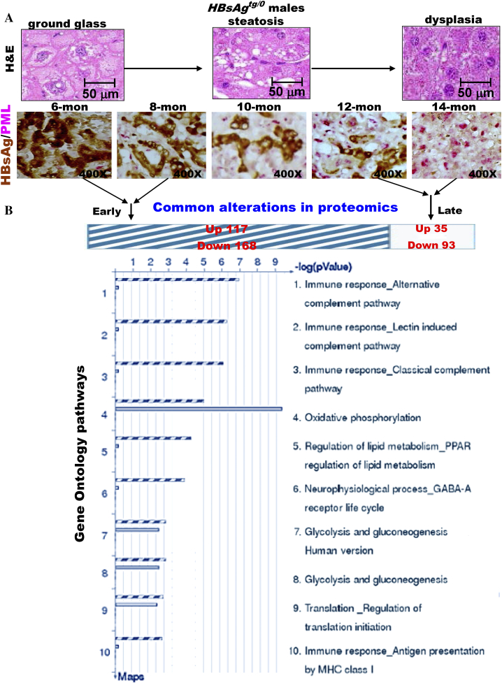
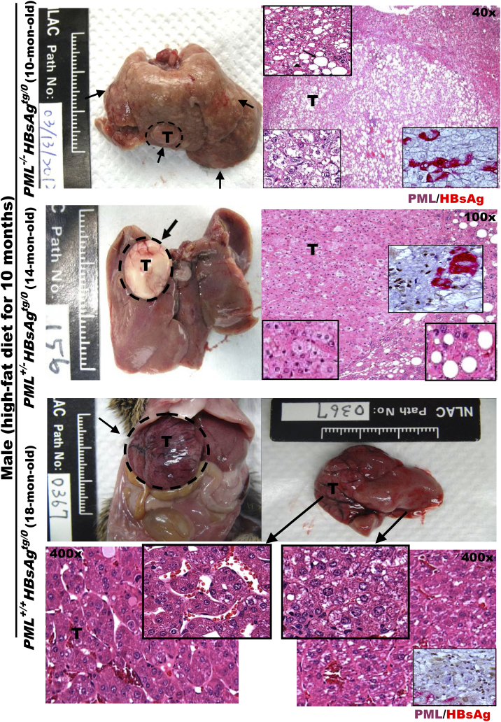
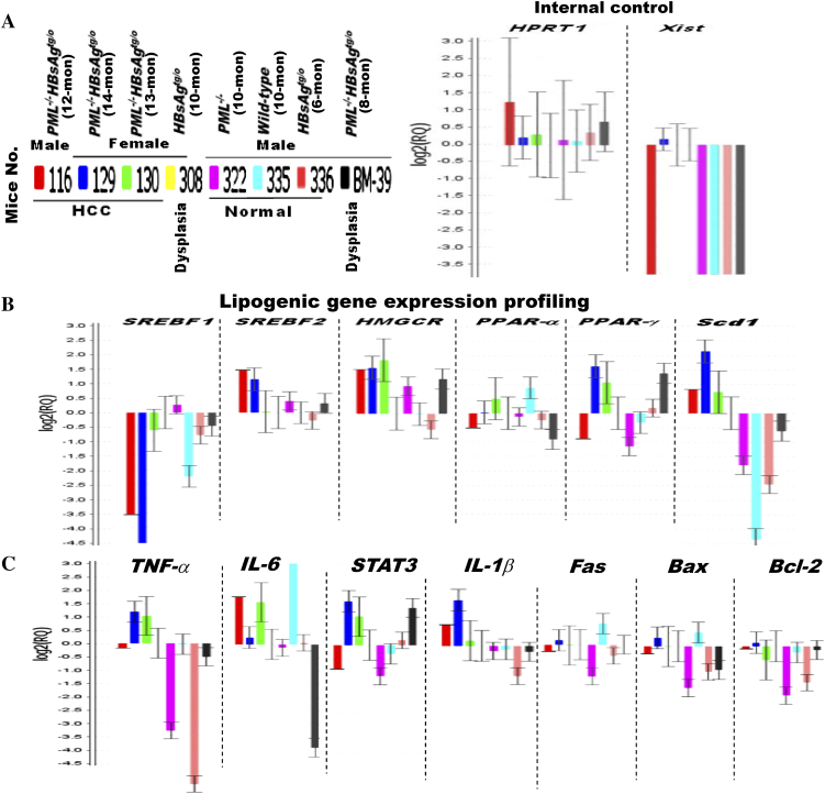
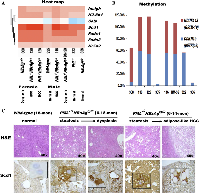
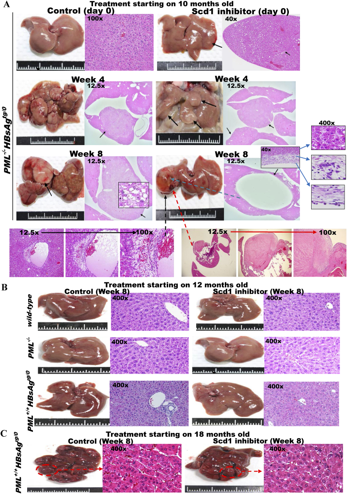
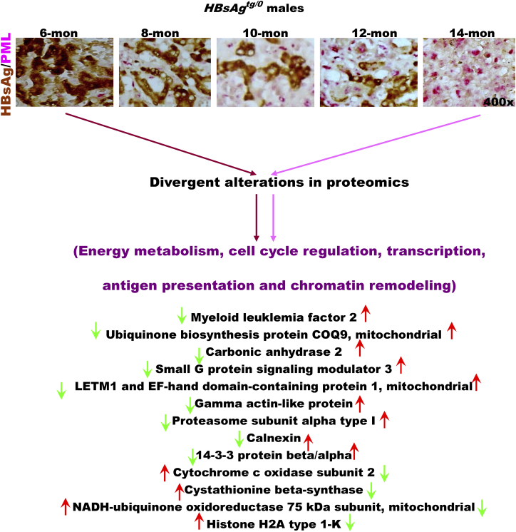
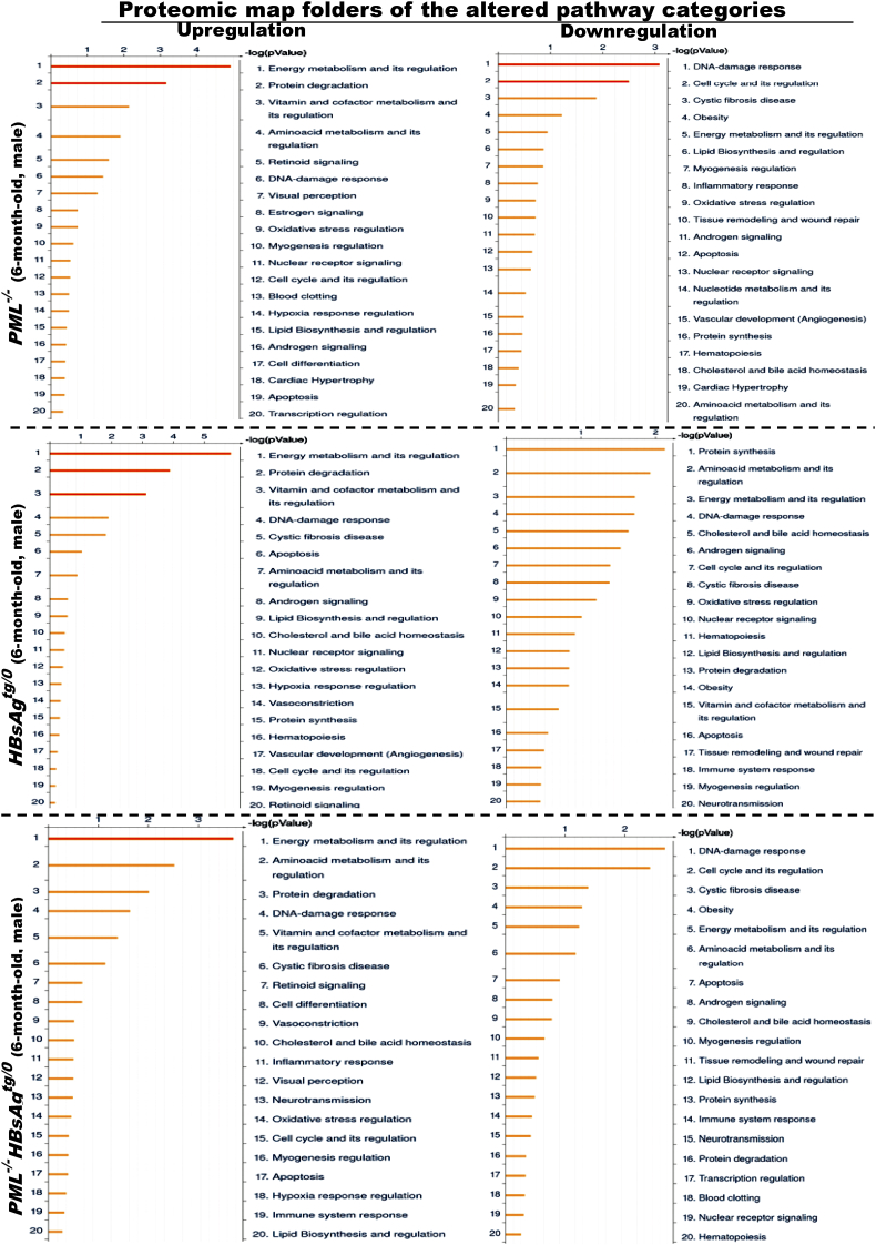
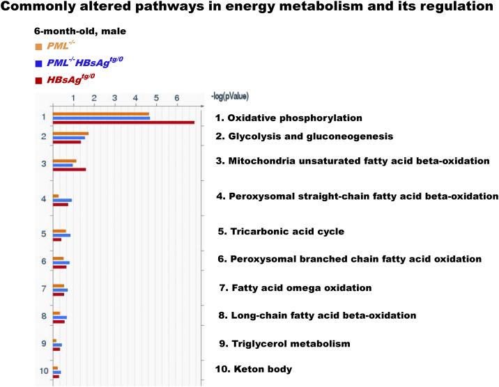
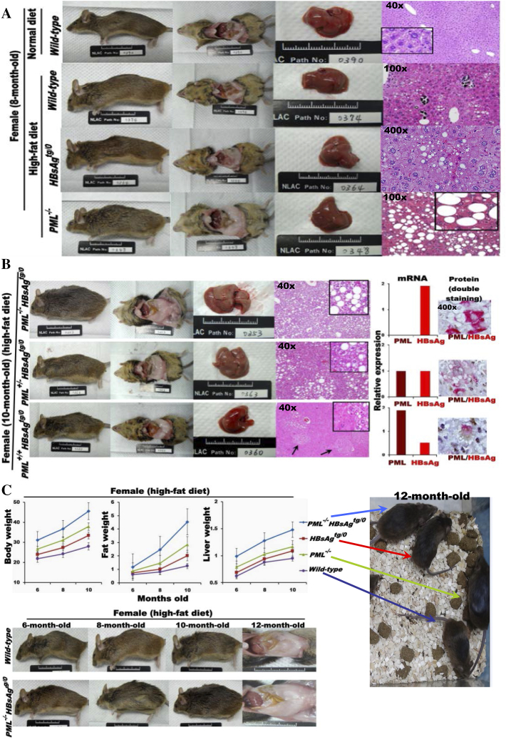
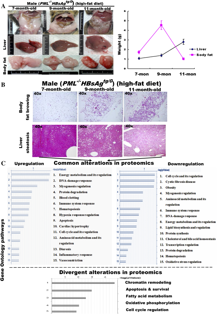
The persistence of hepatitis B surface antigen (HBsAg) is a risk factor for the development of steatosis-associated tumors in chronic hepatitis B virus (HBV) infection, yet little is known about the metabolic link with this factor. We correlated HBV-related pathogenesis in genetically engineered mice and human carriers with metabolic proteomics and lipogenic gene expression profiles. The immunohistochemistry showed that the promyelocytic leukemia protein (PML, a tumor suppressor involved in genome maintenance and fatty acid oxidation), being inversely influenced by the dynamic HBsAg levels from acute phase to seroclearance, appeared as a lipo-metabolic switch linking HBsAg-induced steatosis (lipogenesis) to HBsAg-lost fat-burning hepatocarcinogenesis (lipolysis). Knockdown of PML in HBsAg-transgenic mice predisposed to obesity and drove early steatosis-specific liver tumorigenesis. Proteome analysis revealed that the signaling pathways corresponding to energy metabolism and its regulators were frequently altered by suppression or depletion of PML in the HBsAg-transgenic mice, mainly including oxidative phosphorylation and fatty acid metabolism. Expression profiling further identified upregulation of stearoyl-CoA desaturase 1 (Scd1) and epigenetic methylation of NDUFA13 in the mitochondrial respiratory chain and the cell cycle inhibitor CDKN1c in concordance to the increased severity of lipodystrophy and neoplasia in the livers of HBsAg-transgenic mice with PML insufficiency. The defect in lipolysis in PML-deficient HBsAg-transgenic mice made the HBsAg-induced adipose-like liver tumors vulnerable to synthetic lethality from toxic saturated fat accumulation with a Scd1 inhibitor. Our findings provide mechanistic insights into the evolution of steatosis-associated hepatic tumors driven by reciprocal interactions of HBsAg and PML, and a potential utility of lipid metabolic reprogramming as a treatment target.


Figures

Figure 1
Figure 1
Reciprocal interactions of PML and HBsAg is closely associated with metabolic reprogramming in a chronic HBV mouse model. (A) Representative double-stained images of PML and HBsAg in liver-specific HBsAg-transgenic mice (HBsAgtg/0) at different ages. Note that correlation of the evolving pathology in mice with reciprocal interactions between PML and HBsAg recapitulates the chronic human HBV-infected liver pathogenesis observed in Figure 1C. (B) Comparison of proteomic profiling between young and aged HBsAgtg/0 mice. The enriched genes, whose expression was 2-fold up-regulated or down-regulated in more than 50% of the genetically engineered mice (n = 10 for each genotype) compared to the average expression of the same genes in sex- and age-matched wild-type mice (n = 5 for each group), were categorized by gene ontology (GO) analysis. Note that the major GO pathways affected in young mice with HBsAg-dominant expression and older mice with PML-dominant expression are involved in immune response and energy metabolism, respectively.
Figure 2
Figure 2
Altered PML expression levels reciprocally interacting with HBsAg levels correlate with distinct pathology and phenotypes of lipid metabolism and HBsAg-induced HCCs in mice. Representative images of gross livers and H&E histology with higher magnification of double immunostaining for PML (brown color in the nuclei) and HBsAg (red color in the cytoplasm) from HBsAgtg/0 mice with or without one or two copies of PML knockdown are compared after HCC development. Note that PML−/-HBsAgtg/0, PML+/+HBsAgtg/0 and PML+/-HBsAgtg/0 mice display early-onset adipose-like solid-form HCC, late-onset fatless angiogenic trabecular-type HCC, and mixed adipose-angiogenic features of HCC, respectively. Squares represent in-situ zoomed regions.
Figure 3
Figure 3
Different gene expression profiles of HBsAg-transgenic mice in the presence or absence of PML during HCC development. Comparison of transcriptional profiling in the livers of wild-type, HBsAgtg/0, PML−/− and PML−/-HBsAgtg/0 mice with different ages, sex and pathology. (A) Internal control. (B) Lipogenic genes. (C) Inflammation and apoptosis genes. Error bars indicate mean ± SEM from 3 independent experiments. Note that the up-regulation of Scd1 mRNA levels is in concordance with HCC development in PML−/-HBsAgtg/0 mice.
Figure 4
Figure 4
Correlation of liver pathology with gene expression toward hepatocarcinogenesis in the livers of wild-type, HBsAgtg/0, PML−/− and PML−/-HBsAgtg/0 mice with different ages, sex and pathology. (A) Heat map demonstrating the cluster of differentially expressed genes related to lipid metabolism. Note that Scd1, Fads1 and Fads2 are desaturases involved in fatty acid synthesis and that Nr5a2 regulates cholesterol transport, bile acid homeostasis and steroidogenesis. (B) Epigenetic methylation analysis represented by percentage of growth control genes in livers from the same mice in (A). Note that, in addition to enhanced adipogenesis, PML loss is associated with impairment of the cell cycle and mitochondrial function during HBsAg-induced hepatocarcinogenesis. NDUFA13 (also called GRIM-19), a subunit of the mitochondrial NADH dehydrogenase, functions in the transfer of electrons from NADH to the respiratory chain and as a tumor suppressor by binding to STAT3. CDKN1c (also called p57Kip2) encodes a strong inhibitor of G1 cyclin/Cdk complexes and a negative regulator of cell proliferation. (C) Correlation of liver histology with immunohistochemistry of Scd1 protein expression in HBsAg-transgenic mice with or without PML loss. Note that PML loss induces severe steatosis with diffusely enhanced Scd1 expression and early-onset adipose-like HCC in HBsAg-transgenic mice. Squares represent in-situ zoomed regions.
Figure 5
Figure 5
Scd1 is a metabolic therapeutic target for synthetic lethality in steatosis-associated hepatic tumors of HBsAg-transgenic mice with PML deficiency. Treatment with a small molecule Scd1 inhibitor, A939572, or solution control for HBsAg-transgenic mice with or without PML loss (n = 10–20 for each group). Representative images of gross livers and H&E histology are shown. (A) Ten-month-old PML−/-HBsAgtg/0 mice. Treatment was started when multiple early-onset adipose-like solid-form HCCs (arrows) were developing. Note that the Scd1 inhibitor regressed the HCCs by inducing necrosis of the fatty tumors and caused sinus ectasia with hemorrhage and cyst formation after resolution. (B) Twelve-month-old wild-type, PML−/−and PML+/+HBsAgtg/0 mice. Note that the treatment induced no cytotoxicity in normal liver cells or dysplasia. (C) Eighteen-month-old PML+/+HBsAgtg/0 mice. Note that the late-onset fatless or fat burnt-out angiogenic trabecular-type HCCs (broken circles) did not respond to the Scd1 inhibitor.
Figure 6
Figure 6
PML expression is inversely correlated with dynamic HBsAg levels in human chronic HBV-related pathogenesis. Representative H&E staining and immunostaining of PML (a regulator involved in DNA damage response and repair, cell death and survival, and fatty acid oxidation), HBsAg (an HBV component), Ki-67 (a cell proliferation marker) and/or Scd1 (a key enzyme in fatty acid synthesis) from a normal liver (A), acute HBV infection (B), a chronic HBV carrier (C), and an invasive HBV-related HCC (D). Clinically, chronic HBV pathogenesis can be subdivided into several phases: immune tolerance (high HBsAg levels), immune clearance (decreased HBsAg expression), immune escape (low HBsAg levels), and HCC formation (HBsAg loss). Note that nuclear PML immunoreactivity is strong in acute infection but suppressed in the early phase of chronic infection, which shows intensive cytoplasmic HBsAg staining. PML suppression is relieved upon clearance of HBsAg, indicating a reciprocal interaction between PML and HBsAg. PML restoration appears to correlate with gradually burnt-out steatosis during HCC development while HBsAg is lost. The simultaneously rising PML, Scd1 and Ki-67 immunoactivity in the invasive front of HBsAg-losing HCC cells reflects active fatty acid catabolism and anabolism coupled with cell proliferation. Zoomed regions indicated by arrows are shown in squares.
Figure S1
Figure S1
Divergent alterations in liver proteomics between 6-month-old mice with cytoplasmic ground change and 14-month-old mice with dysplasia in HBsAgtg/0male mice. The enriched genes, whose expression was 2-fold up-regulated or down-regulated in more than 50% of the genetically engineered mice (n = 10 for each genotype) compared to the average expression of the same genes in sex- and age-matched wild-type mice (n = 5 for each group), were categorized by gene ontology (GO) analysis. Note that the divergent expression of proteins is involved in energy metabolism, cell cycle regulation, transcription, antigen presentation and chromatin remodeling.
Figure S2
Figure S2
Liver proteomic map folders of the altered gene ontology pathway categories in 6-month-old PML-/-, HBsAgtg/0and PML-/-HBsAgtg/0male mice. The enriched genes, whose expression was 2-fold up-regulated or down-regulated in more than 50% of the tested mice (n = 10 for each group) compared to the average expression of the same genes in sex- and age-matched wild-type mice (n = 5 for each group), were categorized by gene ontology (GO) pathway analysis. Note that energy metabolism and its regulators are most affected in each group.
Figure S3
Figure S3
The commonly altered metabolic pathways in the liver proteomic map folder of energy metabolism and its regulation. The enriched genes, whose expression was 2-fold up-regulated or down-regulated in more than 50% of the tested mice (n = 10 for each group) compared to the average expression of the same genes in sex- and age-matched wild-type mice (n = 5 for each group), were categorized by gene ontology (GO) pathway analysis. Note that oxidative phosphorylation is the most affected pathway in energy metabolism regulation.
Figure S4
Figure S4
Effects of PML loss or PML suppression on altered lipid metabolism in HBsAg-transgenic mice. In response to challenge with a high-fat (HF) diet, the phenotypic evolution (obesity and body fat mass accumulation) and pathological changes (the severity of steatosis and dysplasia) in the lipid metabolism of the body and liver were compared between HBsAgtg/0 mice with or without one or two copies of PML knockdown (n = 5-10 for each genotype). (A) Respective effects of PML loss and HBsAg accumulation. Squares represent in-situ zoomed regions. (B) Dose effects of PML and HBsAg. Foci of altered hyperplasia and dysplasia are indicated by arrows. Note that PML deficiency induces more steatosis and less dysplasia and that HBsAg stimulation causes more dysplasia and less steatosis. Foci of hyperplasia and dysplasia are indiced by arrows. Squares represent in-situ zoomed regions. (C) Synergistic effects of PML loss and HBsAg accumulation on weight changes in the body, fat and liver. Error bars indicate mean ± SD. Note that PML loss enhances the effects of HBsAg on dysplasia and steatosis and predisposes HBsAgtg/0 mice to body fat mass accumulation and obesity.
Figure S5
Figure S5
PML loss promotes differences in lipid metabolic reprogramming between body fat and the liver in liver-specific HBsAg-transgenic mice.PML-/-HBsAgtg/0 male mice were fed a high-fat diet for 11 months. (A) Weight changes in body fat and liver. Error bars indicate mean ± SD (n = 10-20 for each time point). (B) Representative histology of white body fat mass and liver. Note the continuous fat accumulation in the liver in contrast to the gradual browning and wasting of the body fat. (C) Common and divergent alterations in liver proteomics between 7-month-old and 11-month-old PML-/-HBsAgtg/0 mice. The enriched genes, whose expression was 2-fold up-regulated or down-regulated in more than 50% of the tested mice compared to the average expression of the same genes in sex- and age-matched wild-type mice (n = 5-10 for each group), were categorized by gene ontology (GO) analysis. Note that energy metabolism and cell growth control are the major affected GO pathways.

Similar articles

Cited by

References

    1. Raffetti E, Fattovich G, Donato F. Incidence of hepatocellular carcinoma in untreated subjects with chronic hepatitis B: a systematic review and meta-analysis. Liver Int. 2016;36:1239–1251. - PubMed
    1. Guerrieri F, Belloni L, Pediconi N, Levrero M. Molecular mechanisms of HBV-associated hepatocarcinogenesis. Semin Liver Dis. 2013;33:147–156. - PubMed
    1. Noureddin M, Rinella ME. Nonalcoholic Fatty liver disease, diabetes, obesity, and hepatocellular carcinoma. Clin Liver Dis. 2015;19:361–379. - PMC - PubMed
    1. Karagozian R, Derdák Z, Baffy G. Obesity-associated mechanisms of hepatocarcinogenesis. Metabolism. 2014;63:607–617. - PubMed
    1. Dandri M, Petersen J. Mechanism of Hepatitis B Virus Persistence in Hepatocytes and Its Carcinogenic Potential. Clin Infect Dis. 2016;62(Suppl. 4):S281–8. - PMC - PubMed

LinkOut - more resources